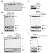
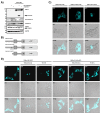
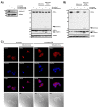
. 2014 May 30;5(10):2988-98.
doi: 10.18632/oncotarget.1634.

Caspase-8-mediated PAR-4 cleavage is required for TNFα-induced apoptosis

Affiliations

Caspase-8-mediated PAR-4 cleavage is required for TNFα-induced apoptosis

Fabian Treude et al. Oncotarget. .

Abstract

The tumor suppressor protein prostate apoptosis response-4 (PAR-4) is silenced in a subset of human cancers and its down-regulation serves as a mechanism for cancer cell survival following chemotherapy. PAR-4 re-expression selectively causes apoptosis in cancer cells but how its pro-apoptotic functions are controlled and executed precisely is currently unknown. We demonstrate here that UV-induced apoptosis results in a rapid caspase-dependent PAR-4 cleavage at EEPD131G, a sequence that was preferentially recognized by caspase-8. To investigate the effect on cell growth for this cleavage event we established stable cell lines that express wild-type-PAR-4 or the caspase cleavage resistant mutant PAR-4 D131G under the control of a doxycycline-inducible promoter. Induction of the wild-type protein but not the mutant interfered with cell proliferation, predominantly through induction of apoptosis. We further demonstrate that TNFα-induced apoptosis leads to caspase-8-dependent PAR-4-cleavage followed by nuclear accumulation of the C-terminal PAR-4 (132-340) fragment, which then induces apoptosis. Taken together, our results indicate that the mechanism by which PAR-4 orchestrates the apoptotic process requires cleavage by caspase-8.


Figures
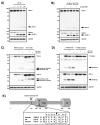
Figure 1
Figure 1. PAR-4 is cleaved during UV-induced apoptosis at position 131 by caspases
(A) HeLa cells were treated with UV (20 mJ × cm−2 at 254 nm) and incubated for the indicated time points. Cell lysates were analyzed by immunoblotting using antibodies against PAR-4, PARP-1 and GAPDH. A processed form recognized by an antibody that preferentially recognizes the N-terminal part of PAR-4 is indicated (cl. PAR-4). (B) HeLa cells were pre-treated with the pan-caspase inhibitor Z-VAD-FMK (20 µM) for 30 min, then treated as in (A) and incubated for the indicated time points. Lysates were analyzed by immunoblotting as above (A). (C) HeLa cells were transiently transfected with the indicated plasmids (1 µg) and after incubation for 24 h treated with UV light as in (A). After further incubation for the indicated time points, cell lysates were analyzed by immunoblotting using two PAR-4 specific antibodies that preferentially recognize the N-terminal part of the protein (upper panel) or the C-terminal part of PAR-4 (middle panel). (D) HeLa cells were transiently transfected with plasmids encoding T7-tagged PAR-4 or T7-tagged PAR-4 D131G (1 µg each). 24 h after transfection and treatment with UV for the indicated times, whole cell lysates were prepared and analyzed by immunoblotting with T7-specific antibodies recognizing the N-terminus of PAR-4 (upper panel) or PAR-4 specific antibodies that recognize the C-terminal part of the protein (middle panel). (E) Schematic overview of human PAR-4 protein: the caspase cleavage site at position 131 adjacent to the SAC domain (Selective for Apoptosis in Cancer cells, amino acids 146-203); NLS sequence (nuclear localisation signal, amino acids 147-163); LZ motif (leucine zipper, amino acids 300-340). The PAR-4 caspase cleavage site is conserved in human and rodent orthologs with the exception of a Ser instead of Gly at position P1' in mouse and rat Par-4.
Figure 2
Figure 2. PAR-4 cleavage leads to inhibition of cell growth
(A) Individual established clones of HeLa Flp-In PAR-4 wild-type (WT), PAR-4 D131G and control (empty vector) T-REx cells were treated with 100 ng/ml doxycycline for 72 h. Protein expression was analyzed by immunoblotting using the indicated antibodies. N-terminal PAR-4 fragments are indicated. (B) For the colony formation assay, 200 cells were seeded and grown for 12 days ± doxycycline prior to staining with methylene blue. Two different clones each for PAR-4 WT, PAR-4 D131G and control cells were analyzed (upper panel). Average values from three independent experiments are displayed. Doxycycline treated cells are shown in light grey. Error bars indicate ± SD. P-value was obtained by two-tailed Student's t-Test, comparing doxycycline treated with non-treated cells (lower panel). (C) Hela Flp-In PAR-4 wild-type and PAR-4 D131G cells were grown for 2 days ± doxycycline. Protein lysates were analyzed with the indicated antibodies.
Figure 3
Figure 3. PAR-4 is a substrate of caspase 8
(A) In vitro caspase assay using FLAG-tagged PAR-4 wild-type and recombinant human caspases 1-10. The assay was stopped after 30 min by adding sample buffer, and the proteins were subjected to immunoblotting using a PAR-4 antibody recognizing the C-terminal part of the protein. (B) HeLa S3 cells were stimulated with TNFα (10 ng/ml) in combination with CHX (0,5 µg/ml) for the indicated time points and with CHX alone for 9 h. Cell lysates were analyzed by immunoblotting with the indicated antibodies and PAR-4 cleavage is indicated. (C) Cell lysates from HeLa S3 control (sh-control) or caspase-8-deficient (sh-caspase-8 #1 and #3) were analyzed by immunoblotting with caspase-8 and GAPDH antibodies (upper panel). HeLa S3 sh-control or sh-caspase-8 #1 and #3 cells were incubated with TNFα (10 ng/ml) in combination with CHX (0,5 µg/ml) for 3 h. Protein lysates were analyzed by protein immunoblotting with antibodies against PAR-4, PARP-1 and GAPDH (lower panel). (D) Caspase-3-deficient MCF-7 cells and MCF-7 cells stably expressing caspase-3 were stimulated with TNFα (10 ng/ml) and CHX (0,5 µg/ml) for the indicated time points and protein lysates were analyzed by immunoblotting with PAR-4, caspase-3 and GAPDH-specific antibodies. PAR-4 cleavage products are indicated. (E) MCF-7 cells were pre-incubated with 50 µM caspase-8 inhibitor (Z-IETD-FMK) for 30 min and subsequently stimulated with TNFα (10 ng/ml) and CHX (0,5 µg/ml) for 16 h. Cell lysates were analyzed with the indicated antibodies via immunoblotting and cleavage of PAR-4 is indicated.
Figure 4
Figure 4. Caspase-8-mediated PAR-4 cleavage promotes apoptosis through nuclear translocation
(A) HEK 293 cells were transiently transfected with plasmids driving expression of PAR-4 wild-type (1 µg), caspase-8 or caspase-3 (50 ng each) for 16 h. Protein lysates were analyzed by protein immunoblotting with the indicated antibodies. PAR-4 cleavage was analyzed with antibodies recognizing the C-terminal part of the protein. (B) Schematic representation of PAR-4 wild-type, PAR-4 D131G and PAR-4 Δ131 fused to eCFP as used for (C). (C) HEK 293 cells were transiently transfected with the plasmids indicated above (B) and after 16 h analyzed by confocal microscopy. DIC: differential interference contrast. (D) HEK 293 cells were transiently transfected as above and after 16 h incubation stimulated with UV (20 mJ × cm−2 at 254 nm) or TNFα (10 ng/ml) and CHX (0,5 µg/ml). 8 h after stimulation cells were analyzed by confocal microscopy.
Figure 5
Figure 5. PAR-4 is required for TNFα-induced apoptosis
(A) Cell lysates from MCF-7 control (sh-control) or caspase-8 deficient (sh-caspase-8 #1 or #3) cells were analyzed by immunoblotting using antibodies against caspase-8 and GAPDH (left panel). MCF-7 control and caspase-8 deficient cells were incubated with CHX (0,5 µg/ml) or TNFα (10 ng/ml) in combination with CHX (0,5 µg/ml) for 10 h. Cell lysates were analyzed by protein immunoblotting with antibodies against PAR-4, PARP-1 and GAPDH and (right panel). N-terminal PAR-4 cleavage products are indicated. (B) MCF-7 control (sh-control) or PAR-4 deficient (sh-PAR-4 #2) cells were treated with CHX (0,5 µg/ml) alone or TNFα (10 ng/ml) in combination with CHX (0,5 µg/ml). After 10 h of incubation cell lysates were analyzed by immunoblotting with antibodies against PAR-4, PARP-1 and GAPDH. N-terminal PAR-4 cleavage products are illustrated. (C) MCF-7 control (sh-control) and caspase-8 deficient cells (sh-caspase-8 #3) were stimulated for 10 h with TNFα (10 ng/ml) and CHX (0,5 µg/ml), immunostained with a C-terminal PAR-4 antibody (red), treated with Hoechst (blue) for nuclear staining and analyzed by confocal microscopy. DIC: differential interference contrast.

References

    1. Sells SF, Wood DP, Jr, Joshi-Barve SS, Muthukumar S, Jacob RJ, Crist SA, Humphreys S, Rangnekar VM. Commonality of the gene programs induced by effectors of apoptosis in androgen- dependent and -independent prostate cells. Cell Growth Differ. 1994;5:457–466. - PubMed
    1. Diaz-Meco MT, Abu-Baker S. The Par-4/PTEN connection in tumor suppression. Cell Cycle. 2009;8:2518–2522. - PMC - PubMed
    1. Cook J, Krishnan S, Ananch S, Sells SF, Shi Y, Walther MM, Linehan WM, Sukhatme VP, Weinstein MH, Rangnekar VM. Decreased expression of the pro-apoptotic protein Par-4 in renal cell carcinoma. Oncogene. 1999;18:1205–1208. - PubMed
    1. Kogel D, Reimertz C, Mech P, Poppe M, Fruhwald MC, Engemann H, Scheidtmann KH, Prehn JH. Dlk/ZIP-kinase-induced apoptosis in human medulloblastoma cells: Requirement of the mitochondrial apoptosis pathway. Br J Cancer. 2001;85:1801–1808. - PMC - PubMed
    1. Moreno-Bueno G, Fernandez-Marcos PJ, Collado M, Tendero MJ, Rodriguez-Pinilla SM, Garcia-Cao I, Hardisson D, Diaz-Meco MT, Moscat J, Serrano M, Palacios J. Inactivation of the candidate tumor suppressor par-4 in endometrial cancer. Cancer Res. 2007;67:1927–1934. - PubMed

Publication types

Substances