Review
. 2014 Jul;24(3):181-91.
doi: 10.1016/j.semradonc.2014.02.007.

Magnetic resonance image guided brachytherapy

Affiliations
Review

Magnetic resonance image guided brachytherapy

Kari Tanderup et al. Semin Radiat Oncol. 2014 Jul.

Abstract

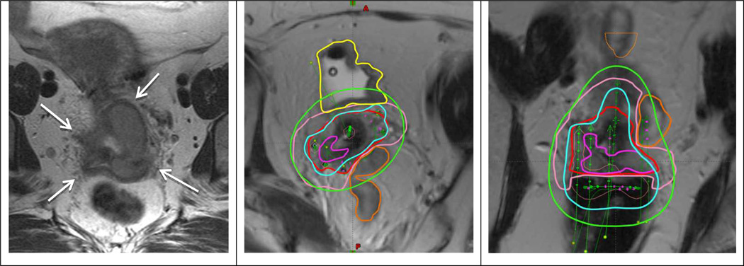
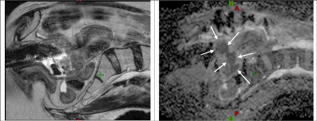
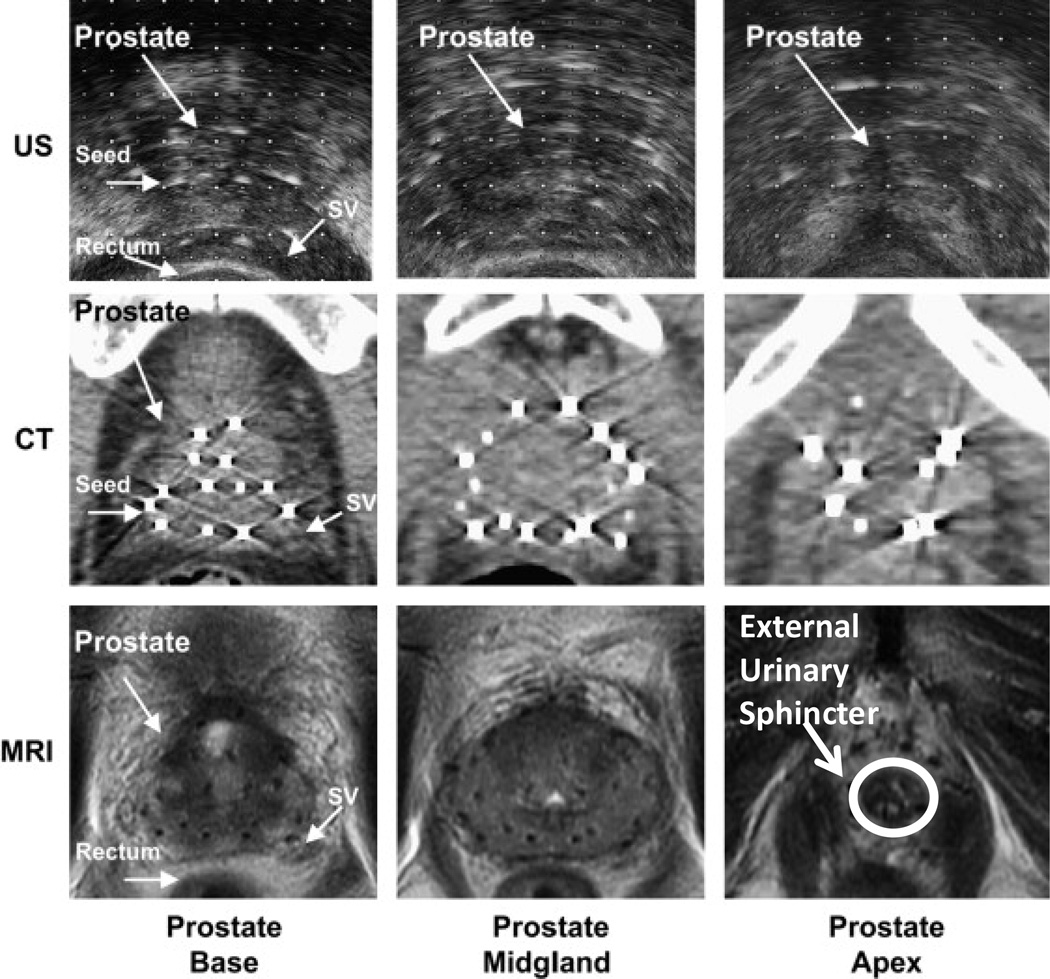
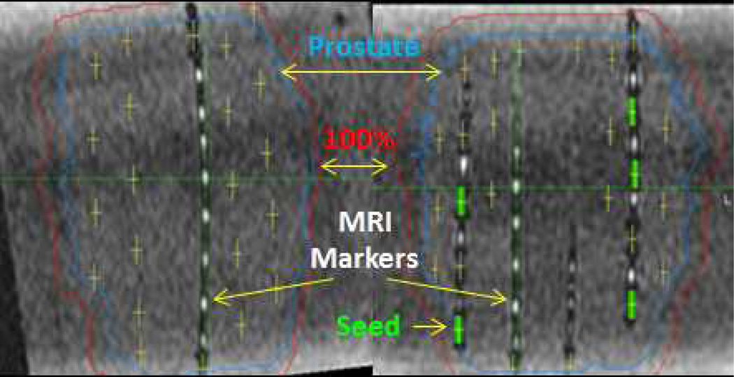
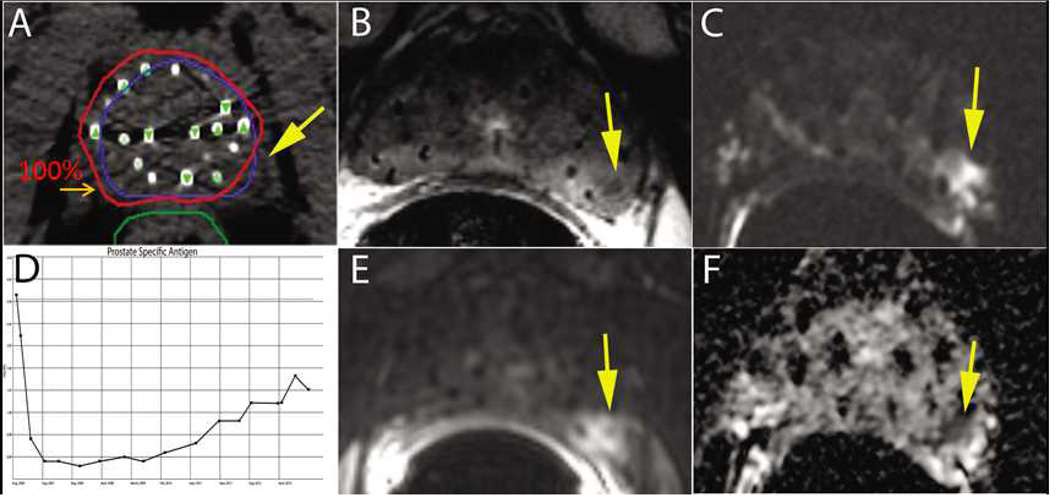
The application of magnetic resonance image (MRI)-guided brachytherapy has demonstrated significant growth during the past 2 decades. Clinical improvements in cervix cancer outcomes have been linked to the application of repeated MRI for identification of residual tumor volumes during radiotherapy. This has changed clinical practice in the direction of individualized dose administration, and resulted in mounting evidence of improved clinical outcome regarding local control, overall survival as well as morbidity. MRI-guided prostate high-dose-rate and low-dose-rate brachytherapies have improved the accuracy of target and organs-at-risk delineation, and the potential exists for improved dose prescription and reporting for the prostate gland and organs at risk. Furthermore, MRI-guided prostate brachytherapy has significant potential to identify prostate subvolumes and dominant lesions to allow for dose administration reflecting the differential risk of recurrence. MRI-guided brachytherapy involves advanced imaging, target concepts, and dose planning. The key issue for safe dissemination and implementation of high-quality MRI-guided brachytherapy is establishment of qualified multidisciplinary teams and strategies for training and education.


Figures

Fig 1
Fig 1
Patient with FIGO stage IIIB treated with EBRT and 2 fractions of PDR MRI-guided brachytherapy. Left panel shows transverse MRI at time of diagnosis with parametrial proximal involvement (left) and to the pelvic wall (right). At time of brachytherapy there was still residual parametrial disease (left proximal and right distal). A combined intracavitary/interstitial applicator (5 needles) was used. Middle and right panel show para-transverse and coronol MRI at time of brachytherapy with the applicator in situ. The volumes are: residual GTV (magenta), CTVHR (red), CTVIR (pink), bladder (yellow) and sigmoid (orange). Isodoses 15 Gy (cyan) and 7.5 Gy (green) correspond to 84 Gy and 60 Gy in terms of total EBRT and brachytherapy EQD2.
Fig 2
Fig 2
MRI at time of brachytherapy for a locally advanced cervical cancer patient with stage IB2 disease treated at Dept of Radiation Oncology, Washington University, St. Louis. Left and right panel show sagittal T2w and ADC images, respectively, obtained at 4. fraction of brachytherapy with the intracavitary applicator in situ. At the time of imaging 13 fractions of IMRT had been delivered with an integrated midline blocking as well as 3 fractions of brachytherapy of 6.5Gy to point A. A significant residual GTV mass is clearly identified in the cervix region on the ADC map (arrows). The bright signal regions on the T2w image indicate residual GTV, but appear with less clear borders towards the surrounding tissue as compared with the ADC map.
Fig 3
Fig 3
Ultrasound (US), CT, and MRI images of the base, midgland, and apex of the prostate following LDR brachytherapy. MRI has superior soft tissue delineation of the prostate over ultrasound and CT. Urinary irritation and bother symptoms, which are more common in prostate brachytherapy, may be reduced with better anatomic delineation of the external urinary sphincter (indicated in bottom right) during simulation and treatment planning. Additionally, better anatomic delineation of the apex, base, neurovascular bundles, bladder neck, and intraprostatic ejaculatory ducts may also improve disease outcomes and reduce treatment related morbidity.
Fig 4
Fig 4
MRI post-implant dosimetry in prostate phantom using C4 MRI markers within strands with I-125 dummy (i.e. non-radioactive) seeds. The positive contrast MRI markers facilitate the localization of the implanted negative contrast I-125 titatium seeds for post-implant MRI-based dosimetry.
Fig 5
Fig 5
A 75 yo male presented with a small nodule at the left base of the gland (i.e. clinical T2a), PSA of 6 ng/ml, and Gleason Score (GS) of 7 (3+4) and was treated with a prostate brachytherapy implant alone to 144 Gy with I-125 (A). Post-implant dosimetry demonstrated a V100=97% and a D90=177 Gy, however, the left midgland was not covered with the 100% isodose line ( A). The patient’s PSA reached a nadir of 0.3 ng/ml eighteen months after the implant. Six years after treatment he had a PSA of 2.3 ng/ml with a PSA doubling time greater than 12 months (D). Multiparametric 1.5T MRI including T2W (B), DWI (C), DCE (E), and ADC maps (F) localized the recurrence at the left midgland. Metastatic workup was negative. Biopsy of the left midgland confirmed adenocarcinoma with a GS of 7 (4+3).

References

    1. Pötter R, Dimopoulos J, Georg P, Lang S, Waldhausl C, Wachter-Gerstner N, et al. Clinical impact of MRI assisted dose volume adaptation and dose escalation in brachytherapy of locally advanced cervix cancer. Radiother Oncol. 2007 May;83(2):148–155. - PubMed
    1. Tanderup K, Nielsen SK, Nyvang GB, Pedersen EM, Rohl L, Aagaard T, et al. From point A to the sculpted pear: MR image guidance significantly improves tumour dose and sparing of organs at risk in brachytherapy of cervical cancer. Radiother Oncol. 2010 Feb;94(2):173–180. - PubMed
    1. Jürgenliemk-Schulz IM, Tersteeg RJ, Roesink JM, Bijmolt S, Nomden CN, Moerland MA, et al. MRI-guided treatment-planning optimisation in intracavitary or combined intracavitary/interstitial PDR brachytherapy using tandem ovoid applicators in locally advanced cervical cancer. Radiother Oncol. 2009 Nov;93(2):322–330. - PubMed
    1. Pötter R, Georg P, Dimopoulos JC, Grimm M, Berger D, Nesvacil N, et al. Clinical outcome of protocol based image (MRI) guided adaptive brachytherapy combined with 3D conformal radiotherapy with or without chemotherapy in patients with locally advanced cervical cancer. Radiother Oncol. 2011 Jul;100(1):116–123. - PMC - PubMed
    1. Lindegaard JC, Fokdal LU, Nielsen SK, Juul-Christensen J, Tanderup K. MRI-guided adaptive radiotherapy in locally advanced cervical cancer from a Nordic perspective. Acta Oncol. 2013 Oct;52(7):1510–1519. - PubMed

Publication types

MeSH terms