. 2014 Jun 19;40(6):924-35.
doi: 10.1016/j.immuni.2014.05.006. Epub 2014 Jun 12.

The parasitophorous vacuole membrane of Toxoplasma gondii is targeted for disruption by ubiquitin-like conjugation systems of autophagy

Affiliations

The parasitophorous vacuole membrane of Toxoplasma gondii is targeted for disruption by ubiquitin-like conjugation systems of autophagy

Jayoung Choi et al. Immunity. .

Abstract

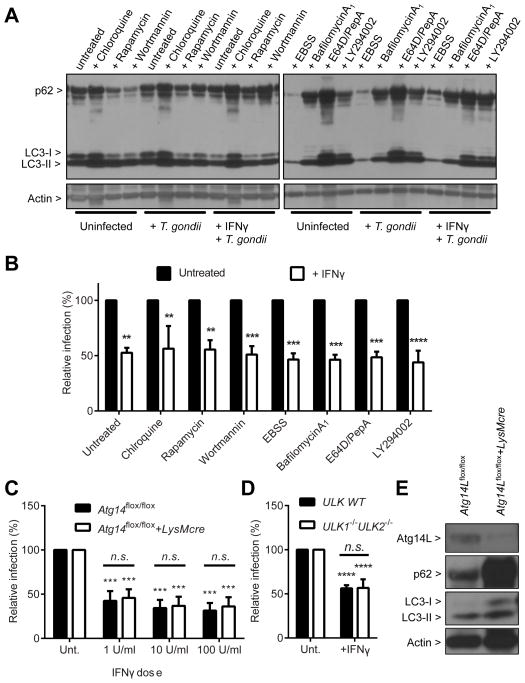
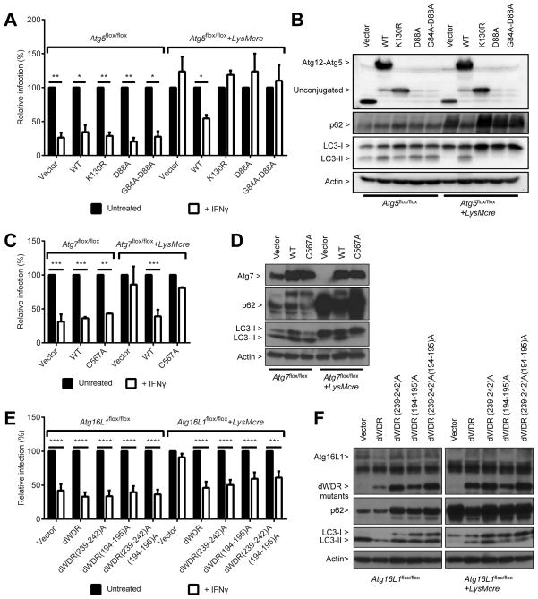
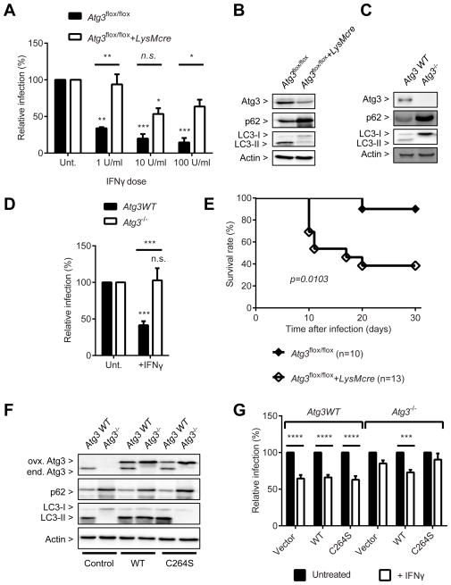
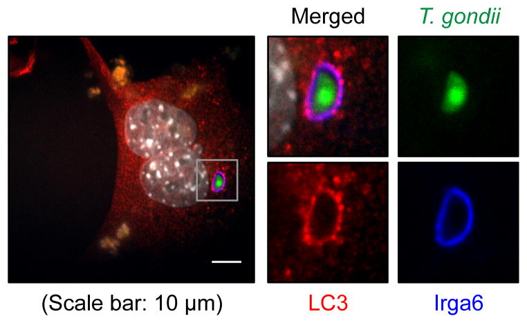
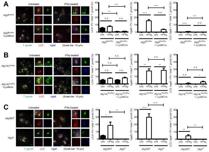
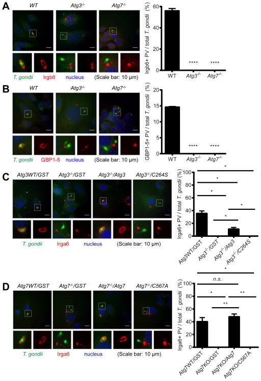
Autophagy is a lysosomal degradation pathway that is important in cellular homeostasis. Prior work showed a key role for the autophagy related 5 (Atg5) in resistance to Toxoplasma gondii. Here we show that the cassette of autophagy proteins involved in the conjugation of microtubule-associated protein 1 light chain 3 (LC3) to phosphatidylethanolamine, including Atg7, Atg3, and the Atg12-Atg5-Atg16L1 complex play crucial roles in the control of T. gondii in vitro and in vivo. In contrast, pharmacologic modulation of the degradative autophagy pathway or genetic deletion of other essential autophagy genes had no substantial effects. Rather the conjugation system was required for targeting of LC3 and interferon-γ effectors onto the vacuolar membrane of T. gondii and its consequent disruption. These data suggest that the ubiquitin-like conjugation systems that reorganize intracellular membranes during canonical autophagy are necessary for proper targeting of immune effectors to the intracellular vacuole membranes utilized by pathogens.


Figures

Figure 1
Figure 1. Atg7 and Atg16L1, but not Atg14L, are required to control T. gondii infection in vivo
Survival curves after intraperitoneal inoculation with 200 (male) or 100 (female) of T. gondii per mouse. Number of mice in parentheses. (A) Atg7flox/flox+/−LysMcre, (B) Atg16L1flox/flox+/−LysMcre, (C) Atg14Lflox/flox+/−LysMcre. Statistical analysis by Log-rank (Mantel-Cox) test. (D) A representative protein blot and quantitative analyses of Atg14L and p62 in the peritoneal exudate macrophages. Combined data as each data point with average. See also Figure S1.
Figure 2
Figure 2. Degradative autophagy pathway is not required for IFNγ to control T. gondii infection
(A) Protein blot for the autophagy status of the samples as shown in (B). Samples harvested at 24 hpi. (B) Flow cytometry analysis for the relative infection (%) of T. gondii at 24 hpi (MOI=1) in BMDMs from C57BL/6 mice treated with chemicals as indicated. Cells were treated with the chemicals for 24 hrs +/− 100 U/ml (50 ng/ml) of IFNγ before infection and treated again during the infection (total 48 hrs of treatment). Autophagy inducers - rapamycin, starvation (EBSS); autophagy inhibitors - wortmannin, LY294002; degradation blockers - chloroquine, bafilomycin A1, E64D, pepstatinA. (C) Same analysis as shown in (B) for Atg14Lflox/flox+/−LysMcre BMDMs. Cells were pretreated with IFNγ for 24 hrs at the indicated doses (D) Same analysis as shown in (B) for WT and Ulk1−/−Ulk2−/− MEFs. (E) Protein blot for untreated/uninfected Atg14Lflox/flox+/−LysMcre BMDMs. Statistical analysis by one-way analysis of variance (1-ANOVA) with Tukey post test. n.s.: not significant (p>0.05), **: p<0.01, ***: p<0.001, ****: p<0.0001. Combined data as average ± SEM. See also Figure S2.
Figure 3
Figure 3. Atg12-Atg5-Atg16L1 complex formation via Atg7 is required for IFNγ to control T. gondii infection
(A) Flow cytometry analysis for T. gondii infection +/− 24 hr pre-treatment of 100 U/ml of IFNγ at 24 hpi (MOI=1) in Atg5flox/flox+/−LysMcre BMDMs transduced with control, WT Atg5 or mutants: K130R - defective in Atg12 conjugation, D88A and G84A/D88A – defective in Atg16L1 binding. (B) Protein blot for untreated or uninfected samples as shown in (A). (C) Same analysis as shown in (A) for Atg7flox/flox+/−LysMcre BMDMs transduced with control, WT Atg7, or enzyme-null mutant (C567A). (D) Protein blot for untreated or uninfected samples as shown in (C). (E) Same analysis as shown in (A) for Atg16L1flox/flox+/−LysMcre BMDMs transduced with control, dWDR (WD repeat deletion), dWDR(239-242)A (+FIP200 binding defective), dWDR(194–195)A (+ mutated 194th–195th), and dWDR(239-242)A(194–195)A. (F) Protein blot for untreated/uninfected samples as shown in (E). Statistical analysis by 1-ANOVA with Tukey post test. *: p < 0.05, **: p<0.01, ***: p<0.001, ****: p<0.0001. Combined data as average ± SEM. See also Figure S3.
Figure 4
Figure 4. Atg3 is required for the control of T. gondii infection
(A) Flow cytometry analysis for T. gondii infection at 24 hpi (MOI=1) in Atg3flox/flox+/−LysMcre BMDMs +/− 24 hr pre-treatment of IFNγ at the indicated doses. (B) Protein blot for untreated or uninfected Atg3flox/flox+/−LysMcre BMDMs. (C) Protein blot for WT and Atg3−/− MEF. (D) Same analysis as shown in (A) for the WT and Atg3−/− MEF. (E) Survival curves of Atg3flox/flox+/−LysMcre mice after intraperitoneal inoculation with T. gondii. (F) Protein blot for untreated or uninfected samples as shown in (G). (G) Same analysis as shown in (A) for WT and Atg3−/− MEFs transduced with control, WT Atg3, or its enzyme-null mutant (C264S) +/− 24 hr pre-treatment of 100 U/ml of IFNγ. Statistical analysis by 1-ANOVA with Tukey post test or Log-rank (Mantel-Cox) test. n.s.: not significant (p>0.05), **: p<0.01, ***: p<0.001, ****: p<0.0001. Combined data as average ± SEM. See also Figure S3.
Figure 5
Figure 5. LC3 and Irga6 localize on the PVM of T. gondii
(A) A representative image of immunofluorescence for WT MEF at 2 hpi (MOI=1) of T. gondii infection with 24 hr pre-treatment of 100 U/ml of IFNγ. Experiments were performed more than thrice.
Figure 6
Figure 6. Atg5 and Atg3, but not Atg14L, are required for the localization of LC3 and Irga6 on the PVM of T. gondii
Representative images (left) and quantitation (right) of immunofluorescence for T. gondii, LC3, and Irga6 in (A) Atg5flox/flox+/−LysMcre and (B) Atg14Lflox/flox+/−LysMcre BMDMs and (C) Atg3 WT and Atg3−/− MEFs at 2 hpi (MOI=1) of T. gondii infection +/− 24 hr pre-treatment of 100 U/ml of IFNγ. At least 100 cells infected T. gondii were analyzed for quantitation. Statistical analysis by 1-ANOVA with Tukey post test. n.s.: not significant (p>0.05), **: p<0.01, ***: p<0.001, ****: p<0.0001. Combined data as average ± SEM.
Figure 7
Figure 7. The ubiquitin-like conjugation systems of the autophagy pathway are required for the localization of IFNγ effectors onto the PVM of T. gondii infection
Immunofluorescence of MEFs at 2 hpi (MOI=1) of T. gondii infection with 24 hr pre-treatment of 100 U/ml of IFNγ. Representative images (left) and quantitation (right) of immunofluorescence. Irgb6 (A) and GBP1-5 (B) in WT, Atg3−/− and Atg7−/− MEFs. At least 50 of infected T. gondii were analyzed for quantitation. Statistical analysis by 1-ANOVA with Tukey post test. ****: p < 0.0001. Experiments were performed twice. Combined data as average ± SEM. (C) T. gondii and Irga6 in Atg3 WT and Atg3−/− MEFs transduced with control (GST, Glutathione-S-transferase), WT Atg3, or enzyme-null mutant (C264S). (D) T. gondii and Irga6 in Atg7 WT and Atg7−/− MEFs transduced with control (GST), WT Atg7, or enzyme-null mutant (C567A). At least 100 of infected T. gondii were analyzed for quantitation. Statistical analysis by unpaired t-test. n.s.: not significant. *: p < 0.05, **: p < 0.01. Experiments were performed twice. Combined data as average ± SEM. See also Figure S4.

References

    1. Bestebroer J, V’kovski P, Mauthe M, Reggiori F. Hidden behind autophagy: the unconventional roles of ATG proteins. Traffic. 2013;14:1029–41. - PMC - PubMed
    1. DeSelm CJ, Miller BC, Zou W, Beatty WL, van Meel E, Takahata Y, Klumperman J, Tooze SA, Teitelbaum SL, Virgin HW. Autophagy proteins regulate the secretory component of osteoclastic bone resorption. Dev Cell. 2011;21:966–74. - PMC - PubMed
    1. Florey O, Kim SE, Sandoval CP, Haynes CM, Overholtzer M. Autophagy machinery mediates macroendocytic processing and entotic cell death by targeting single membranes. Nat Cell Biol. 2011;13:1335–43. - PMC - PubMed
    1. Fujita N, Morita E, Itoh T, Tanaka A, Nakaoka M, Osada Y, Umemoto T, Saitoh T, Nakatogawa H, Kobayashi S, et al. Recruitment of the autophagic machinery to endosomes during infection is mediated by ubiquitin. J Cell Biol. 2013;203:115–28. - PMC - PubMed
    1. Fujita N, Itoh T, Omori H, Fukuda M, Noda T, Yoshimori T. The Atg16L complex specifies the site of LC3 lipidation for membrane biogenesis in autophagy. Mol Biol Cell. 2008;19:2092–100. - PMC - PubMed

Publication types

MeSH terms