. 2014 Jun 19;40(6):936-48.
doi: 10.1016/j.immuni.2014.05.007. Epub 2014 Jun 12.

Antiviral activity of human OASL protein is mediated by enhancing signaling of the RIG-I RNA sensor

Affiliations

Antiviral activity of human OASL protein is mediated by enhancing signaling of the RIG-I RNA sensor

Jianzhong Zhu et al. Immunity. .

Abstract

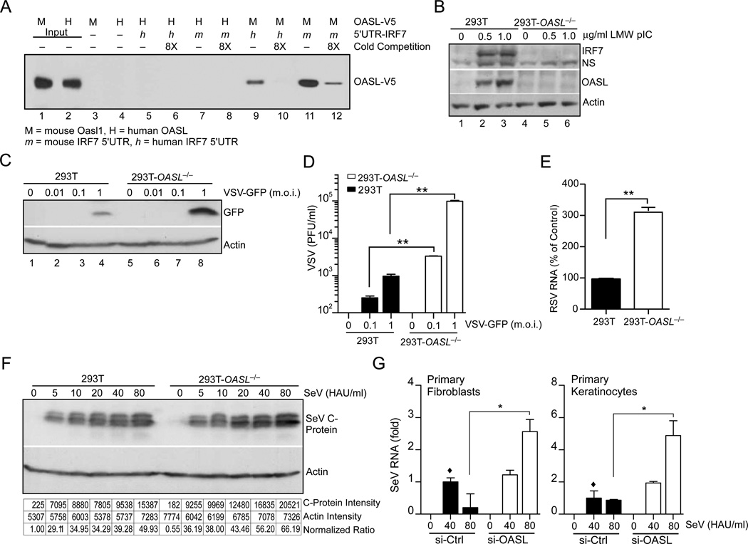
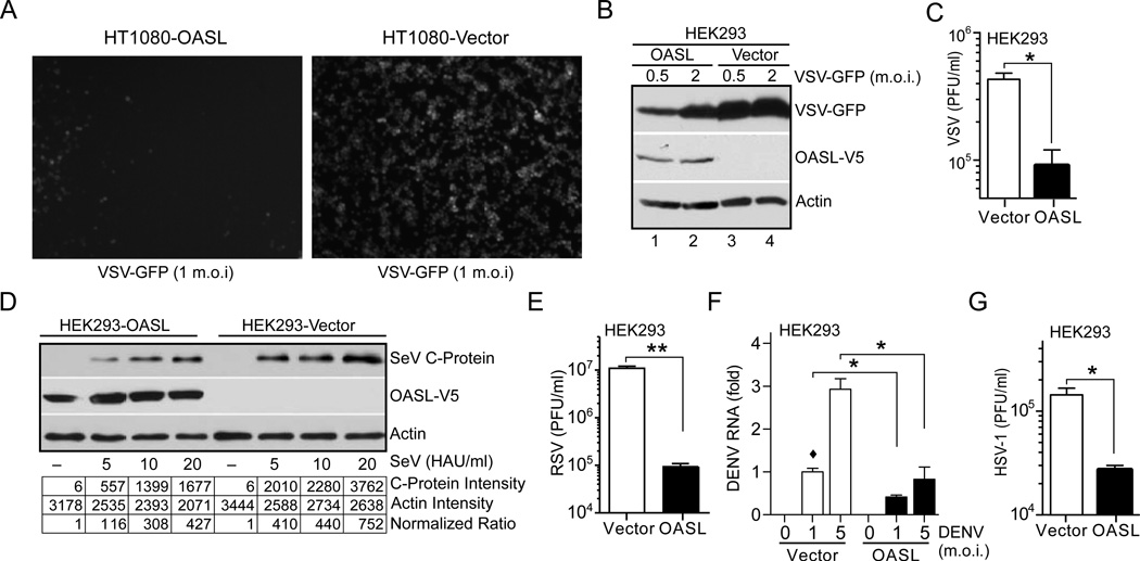
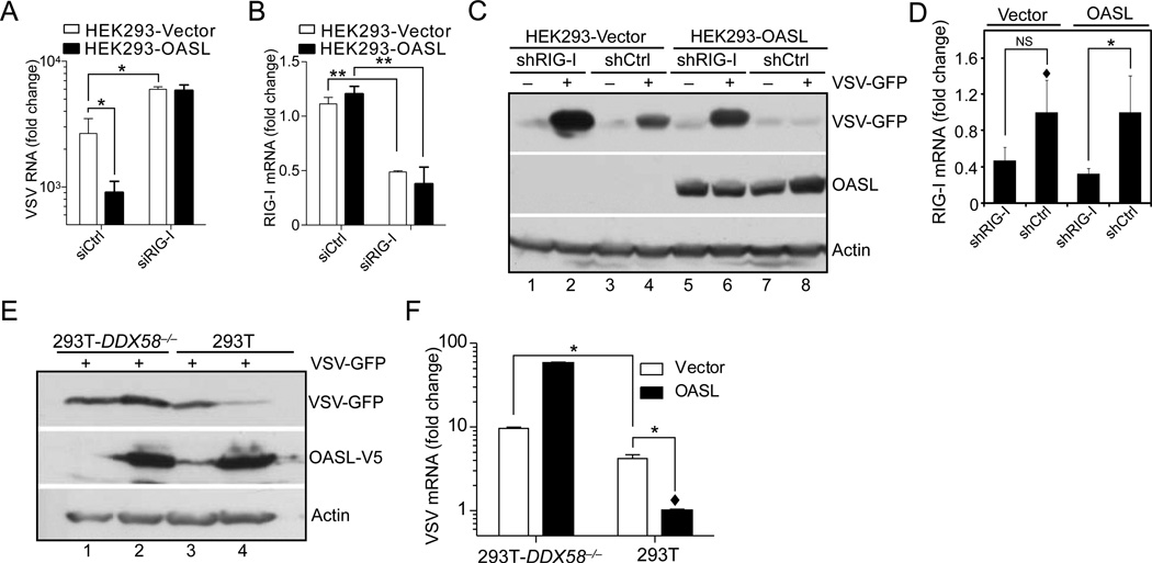
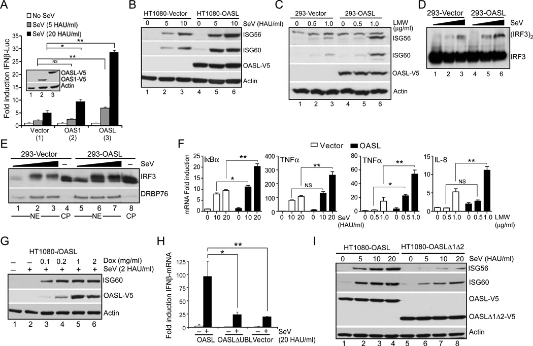
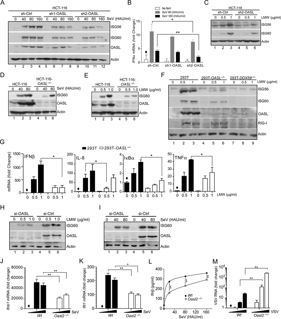
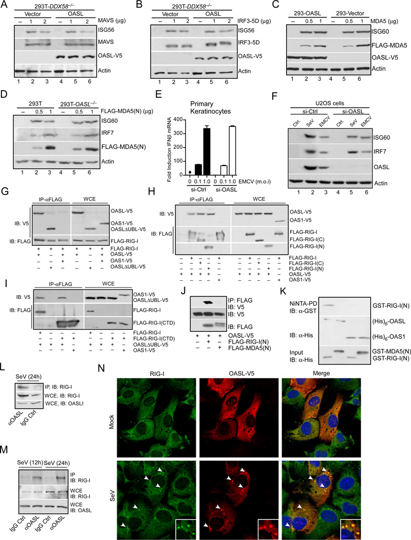
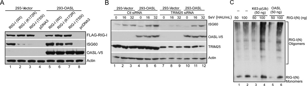
Virus infection is sensed in the cytoplasm by retinoic acid-inducible gene I (RIG-I, also known as DDX58), which requires RNA and polyubiquitin binding to induce type I interferon (IFN) and activate cellular innate immunity. We show that the human IFN-inducible oligoadenylate synthetases-like (OASL) protein has antiviral activity and mediates RIG-I activation by mimicking polyubiquitin. Loss of OASL expression reduced RIG-I signaling and enhanced virus replication in human cells. Conversely, OASL expression suppressed replication of a number of viruses in a RIG-I-dependent manner and enhanced RIG-I-mediated IFN induction. OASL interacted and colocalized with RIG-I, and through its C-terminal ubiquitin-like domain specifically enhanced RIG-I signaling. Bone-marrow-derived macrophages from mice deficient for Oasl2 showed that among the two mouse orthologs of human OASL, Oasl2 is functionally similar to human OASL. Our findings show a mechanism by which human OASL contributes to host antiviral responses by enhancing RIG-I activation.


Figures

Fig. 1
Fig. 1. Unlike mouse Oasl1, human OASL does not bind IRF7 mRNA, and its loss enhances replication of a number of RNA viruses
(A) Comparison between human and mouse IRF7 5’-UTR binding to human OASL or mouse Oasl1 proteins. Biotin labeled IRF7 5’-UTR RNA were incubated with the partially purified V5 tagged OASL or Oasl proteins and pulled down by streptavidin beads. Following extensive wash, bead-bound proteins were analyzed by immunoblotting (IB) using V5 antibody. (B) Reduced IRF7 induction in 293T-OASL−/− cells. 293T-OASL−/− and control 293T cells were transfected with LMW for 24 h, and analyzed by IB. NS, Non-specific. (C–D) VSV replication in 293T-OASL−/− cells detected by IB using anti-GFP antibody (C) or by plaque assay (D) 24 h post infection. (E) RSV replication in 293T-OASL−/− cells. Cells were infected with RSV at 3 m.o.i. for 18 h followed by total RNA extraction and detection by qRT-PCR. (F) SeV replication in 293T-OASL−/− cells detected by IB using SeV C protein antibody 24 h post infection. Densitometric analysis of the immunoblot is shown below. (G) OASL silencing results in higher SeV replication in primary human cells. Primary human foreskin fibroblasts or keratinocytes were transfected using lipfectamine RNAi MAX with either 50 nM OASL siRNA or control siRNA (ON-TARGETplus, SMARTpool, Thermo Scientific) for 48 h. The cells were then infected with SeV at indicated doses for another 24 h followed by detection of SeV specific RNA by qRT-PCR.
Fig. 2
Fig. 2. OASL expression provides cellular antiviral activity
(A) OASL expression inhibits VSV infection. Cells were infected with VSV-GFP at 1 m.o.i. GFP florescence was observed under florescence microscope 8 h post-infection. Representative micrograph from at least three separate experiments is shown. (B–C) Expression of OASL reduces VSV replication. Cells were infected with VSV-GFP at the indicated m.o.i for 8 h followed by IB using GFP antibody (B). Supernatants from similarly infected cells (5 m.o.i) were used for plaque assay on BHK21 cells (C). (D) OASL expression inhibits SeV infection. SeV infected (24 h) cells were analyzed by IB using antibody against SeV C protein. Densitometric analysis of the protein bands are shown below. (E–G) OASL expression inhibits RSV, DENV and HSV-1 replication. Cells were infected with RSV (3 m.o.i) for 48 h, followed by plaque assay on Hep2 cells (E). Cells were infected with type 2 dengue virus (DENV) at indicated doses for 48 h followed by qRT-PCR for DENV specific RNA (F). Cells were infected with HSV-1 at 5 m.o.i. for 24 h followed by plaque assay on Vero cells (G).
Fig. 3
Fig. 3. OASL antiviral activity is dependent on RIG-I
(A–B) Loss of RIG-I expression reduces OASL antiviral activity against VSV. Cells were transfected with 50 nM RIG-I or control siRNA (Dharmacon) for 48 h, followed by VSV infection (1 m.o.i., 24 h). Total RNA from the infected cells were extracted and analyzed for VSV replication (A) and RIG-I mRNA (B) by qRT-PCR. (C) Indicated cells were transfected with psiRNA-Ctrl or psiRNA-hRIG-I plasmids (Invivogene) followed by Zeocin selection. VSV infected (1 m.o.i) cells were immunobloted with indicated antibodies. (D) Levels of RIG-I silencing were estimated by qRT-PCR. (E–F) Expression of OASL in RIG-I null cells does not protect them from VSV. OASL expressing 293T-DDX58−/− and control cells were infected with 1 m.o.i VSV for 24 h followed by IB with indicated antibodies (E), and qRT-PCR to detect VSV replication (F).
Fig. 4
Fig. 4. OASL enhances RIG-I signaling
(A) OASL expression enhances IFNβ-reporter activity. HEK293 cells were co-transfected either with control pcDNA3 (1), pcDNA3-OAS1 (2) or pcDNA3-OASL (3) in combination with IFNβ-luciferase reporter and β-actin Renilla luciferase reporter for 24 h. One part of the transfected cells was used for IB to detect protein expression (inset) and the other part was stimulated with SeV for 16 h and luciferase activities were measured. (B–C) OASL expression enhances RIG-I signaling. Cells were infected with SeV for 16 h followed by IB using ISG56, ISG60, V5 (OASL) and actin antibodies (B). Cells were transfected with LMW for 24 h followed by IB using indicated antibodies (C). (D–E) IRF3 dimerization and nuclear localization is enhanced in OASL expressing cells. Indicated cells were stimulated with SeV for 12h. Cell lysates were analyzed in native-PAGE followed by IB with IRF3 antibody (D). Nuclear extracts (NE) from similarly stimulated cells were analyzed along with control uninfected cytoplasm (CP) by IB for IRF3 and nuclear marker DRBP76 respectively (E). (F) NF-κB-dependent gene inductions are also enhanced in OASL expressing cells. Indicated cells were either transfected with LMW or infected with SeV for 24 h followed by qRT-PCR analysis using indicated primers to detect NF-κB-controlled gene induction. (G) Dose-dependent enhancement of RIG-I-signaling sensitivity by OASL. HT1080-iOASL cells were stimulated with Doxycycline at indicated concentrations for 48h followed by infection with SeV (2 HAU/ml) for 8 h. Cell lysates were analyzed by IB as indicated. (H) UBL deleted OASL mutant does not enhance SeV mediated IFNβ induction. Cells were either mock infected or infected with 20 HAU/ml SeV for 16 h. Total RNA from each well were extracted followed by qRT-PCR using IFNβ specific primers. (I) OASL mediated enhancement of ISG induction by SeV is dependent on its UBL. Various OASL expressing HT1080 cells were infected with SeV as indicated for 16 h followed by IB with indicated antibodies as before.
Fig. 5
Fig. 5. Loss of human OASL and mouse Oasl2 reduces RIG-I signaling and enhances virus replication
(A–C) Inhibition of RIG-I signaling in HCT-116 cells following OASL silencing. HCT-116 cells stably expressing OASL shRNA (two different shRNA, sh1-OASL and sh2-OASL targeting two different regions of OASL mRNA) or control vector stimulated with SeV as indicated for 24h followed by IB with indicated antibodies (A). Indicated cells were stimulated with SeV as in (A), and the cellular RNA were analyzed for IFNα mRNA by qRT-PCR (B). Same HCT-116 OASL shRNA and control cells were transfected with LMW for 24 h followed by IB with the indicated antibodies (C). (D–E) Reduction of RIG-I-mediated ISG induction in HCT-116-OASL−/− cells. OASL−/− HCT-116 and wild type HCT-116 cells were either infected with SeV (D) or transfected with LMW (E) as indicated, followed by IB for ISG60, OASL and actin antibodies respectively. (F) ISG induction in OASL and RIG-I targeted 293T cells. Wild type 293T, 293T-OASL−/− and 293T-DDX58−/− cells were transfected with LMW for 24 h, followed by IB for the indicated proteins. (G) Effect of OASL loss on RIG-I-mediated induction of NF-κB target genes. Cells were treated similarly as in (F) followed by qRT-PCR analysis of indicated NF-κB-regulated genes. (H–I) OASL silencing reduces ISG60 induction by RIG-I signaling in primary human keratinocytes. Primary keratinocytes were transfected either with OASL or control siRNA for 48 h, followed by either transfection with LMW or infection with SeV at the indicated doses for another 24 h and analysis by IB. (J–K) Downregulation of RIG-I-mediated Ifnb1 (IFNβ) and Ifit1 (ISG56) mRNA induction in Oasl2−/− macrophages. BMDM from wild type (Wt) or Oasl2−/− mice were infected with SeV for 8 h followed by detection of Ifnb1 (J) and Ifit1 (K) mRNA by qRT-PCR. (L) Secreted IFNβ was measured in the supernatants from SeV infected (8h) Wt (●) and Oasl2−/− (○) BMDM using the mouse IFNβ ELISA kit. (M) Increased VSV replication in BMDM from Oasl2−/− mice. BMDM from Wt or Oasl2−/− mice were infected with VSV for 24 h followed by quantitation of VSV RNA by qRT-PCR.
Fig. 6
Fig. 6. OASL binds to and specifically enhances RIG-I signaling
(A–B) OASL does not affect RLR signaling downstream of MAVS. 293T-DDX58−/− cells stably expressing OASL or vector control were transfected with indicated plasmids to express either MAVS (A) or constitutively active IRF3-5D (B) for 24 h followed by immunoblot analysis. (C) OASL does not enhance MDA5 signaling. Cells were transfected either with vector or two different concentrations of FLAG-MDA5 plasmid for 24 h followed by IB. (D) Loss of OASL expression does not affect MDA5 signaling. Cells were transfected for 24 h with indicated amounts of MDA5(N) plasmid and analyzed by IB. (E) OASL silencing does not affect IFNβ induction stimulated by EMCV infection in keratinocytes. Primary human keratinocytes were transfected with 50 nM OASL siRNA and control siRNA for 48 h, followed by EMCV infection for another 24 h. The OASL silencing (Suppl. Fig. S1E), IFNβ production (E) and EMCV replication (Suppl. Fig. S1E) were analyzed by specific qRT-PCR. (F) OASL does not affect the gene ISG60 and IRF7 induction triggered by a physiological MDA5 agonist. 293T cells were infected either with EMCV, VSV and SeV or mock infected (Ctrl.) and extracted for total RNA as described before (Jiang et al., 2012). U2OS cells were first transfected with control or OASL siRNA as described in Fig. 1 for 48 h followed by transfection with 1mg of total RNA prepared form infected cells as indicated. Cell lysates were analyzed by IB. (G) RIG-I interacts with OASL. 293T cells were co-transfected for 24 h with FLAG-RIG-I and OASL-V5, OASL-ΔUBL-V5 or OAS1-V5 plasmids, immunoprecipitated (IP) with FLAG antibody, followed by IB with V5 antibody. Whole cell extracts (WCE) from transfected cells were similarly analyzed for expression levels. (H) Both N-terminal CARD and C-terminal Helicase-CTD domains of RIG-I interact with OASL. 293T cells were either individually transfected with OASL-V5 or co-transfected as before with indicated plasmids followed by IP analysis. (I) OAS domain of OASL interacts with RIG-I CTD domain. 293T cells were transfected as indicated, followed by similar IP analysis. (J) RIG-I CARD domain (RIG-I(N)) specifically interacts with OASL, while MDA5 CARD domain (MDA5(N)) does not. 293T cells were transfected as indicated followed by IP analysis. (K) Specific in vitro interaction of RIG-I(N) with OASL. GST-RIG-I(N) and GST-MDA5(N) proteins purified from bacteria were incubated on ice as indicated with (His)6-OASL or with (His)6-OAS1 purified from insect cells followed by Ni-NTA pull down and IB. (L) Interaction of endogenous OASL and RIG-I. HEK293 cells were infected with SeV (100 HAU/ml) for 24 h. Cell lysates were immunoprecipitated either with OASL antibody or control IgG followed by IB with RIG-I antibody. Control WCE was analyzed to show expression. (M) Interaction of endogenous RIG-I and OASL in primary human fibroblsts. Human primary foreskin fibroblasts (HFF) were stimulated with SeV (160 HAU/ml) for 12 and 24 h respectively, followed by IP analysis as indicated. (N) OASL co-localizes with RIG-I in virus infected cells. HT1080-OASL cells were either mock infected or infected with 100 HAU/ml SeV for 8 h immuno-stained with RIG-I and V5 antibodies followed by confocal microscopy. The arrows denote the puntae formation, and insets show magnified puntaes.
Fig. 7
Fig. 7. OASL specifically enhances RIG-I-signaling by mimicking pUb to activate RIG-I
(A) Ubiuitination or p(Ub) binding mutants of RIG-I can be activated in presence of OASL. Cells were transfected with wild type (Wt) or mutant RIG-I constructs for 24 h and analyzed by IB with indicated antibodies. (B) OASL is capable of promoting RIG-I activation in the absence of TRIM25. Cells were transfected with 50 nM TRIM25 siRNA (Zeng et al., 2010) or control siRNA for 48 h followed by SeV infection for 16 h and IB analysis. (C) OASL enhances RIG-I(N) oligomerizaiton similar to pUb. 50 or 100 ng of purified RIG-I(N) (GST tagged) were incubated either with K63-linked polyubiquitin chain (mixture of (pUb)>3, Boston Biochem) or with purified OASL for 1h at 30°C in the binding buffer (20 mM HEPES-KOH pH 7.4; 5 mM MgCl2 and 1X proteinase inhibitor cocktails). The incubated proteins were analyzed by native PAGE followed by IB with anti-GST antibody.

References

    1. Ablasser A, Bauernfeind F, Hartmann G, Latz E, Fitzgerald KA, Hornung V. RIG-I-dependent sensing of poly(dA:dT) through the induction of an RNA polymerase III-transcribed RNA intermediate. Nat Immunol. 2009;10:1065–1072. - PMC - PubMed
    1. Bowie AG, Unterholzner L. Viral evasion and subversion of pattern-recognition receptor signalling. Nat Rev Immunol. 2008;8:911–922. - PMC - PubMed
    1. Chiu YH, Macmillan JB, Chen ZJ. RNA polymerase III detects cytosolic DNA and induces type I interferons through the RIG-I pathway. Cell. 2009;138:576–591. - PMC - PubMed
    1. Civril F, Bennett M, Moldt M, Deimling T, Witte G, Schiesser S, Carell T, Hopfner KP. The RIG-I ATPase domain structure reveals insights into ATP-dependent antiviral signalling. EMBO Rep. 2011;12:1127–1134. - PMC - PubMed
    1. Eskildsen S, Justesen J, Schierup MH, Hartmann R. Characterization of the 2'-5'-oligoadenylate synthetase ubiquitin-like family. Nucleic acids research. 2003;31:3166–3173. - PMC - PubMed

Publication types

MeSH terms