

The cervical myodural bridge, a review of literature and clinical implications
- PMID: 24932022
- PMCID: PMC4025088
The cervical myodural bridge, a review of literature and clinical implications
Abstract
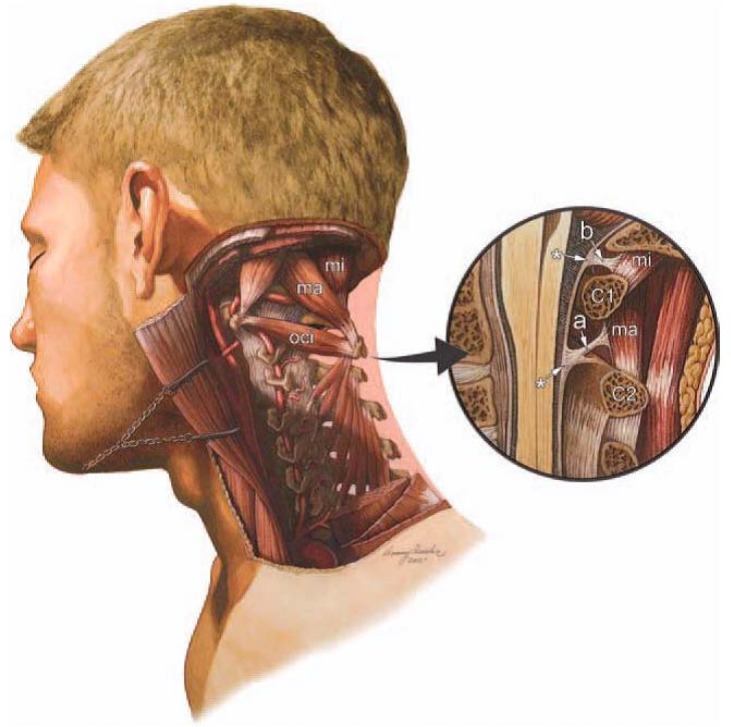
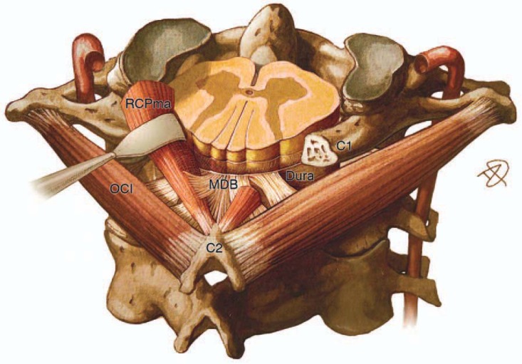
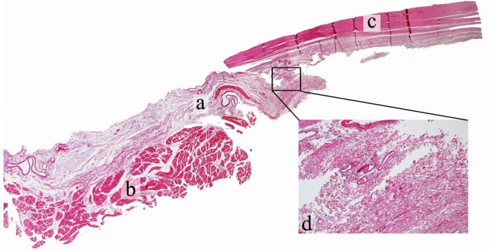
The role of posterior cervical musculature in sensorimotor control, cervicocephalic pain, and stabilization of the spinal cord has been recently described. Anatomical soft tissue connections which cross the cervical epidural space link suboccipital muscle fascia and dura. These myodural bridges provide passive and active anchoring of the spinal cord. They may also be involved in a dural tension monitoring system to prevent dural infolding, and maintain patency of the spinal cord. Modulation of dural tension may be initiated via a sensory reflex to muscular contractile tissues. Unanticipated movements such as hyperflexion extension injuries stimulate deep suboccipital muscles and transmit tensile forces through the bridge to the cervical dura. Due to its larger cross sectional area, the rectus capitis posterior major myodural bridge may exert greater mechanical traction on the dura than the rectus capitis posterior minor. University ethics committee approval and anatomical donor consent was obtained for this study.
Le rôle de la musculature cervicale postérieure dans le contrôle sensorimoteur, la douleur cervico-céphalique et la stabilisation de la moelle épinière n’a que récemment fait l’objet d’une description. Les connexions anatomiques des tissus mous qui traversent l’espace épidural cervical lient le fascia et la dure-mère des muscles sous-occipitaux. Ces ponts myoduraux offrent un point d’ancrage passif et actif à la moelle épinière. Ils peuvent aussi participer au système de contrôle de la tension durale afin de prévenir le repliement dural et de maintenir la perméabilité de la moelle épinière. Les modulations de la tension durale peuvent être provoquées par un réflexe sensoriel aux tissus musculaires contractiles. Les mouvements non anticipés comme les blessures résultant d’une hyperflexion-extension stimulent les muscles sous-occipitaux profonds et transmettent des efforts de traction par le pont sur la dure-mère cervicale. En raison de sa plus grande section transversale, le pont myodural grand droit postérieur peut exercer une plus grande traction mécanique sur la dure-mère que le muscle petit droit postérieur. L’approbation du comité d’éthique de l’université et le consentement du donneur anatomique ont été obtenus pour la présente étude.
Keywords: dura mater; myodural bridge; obliquus capitis inferior; rectus capitis posterior major.
Figures
Similar articles
-
The obliquus capitis inferior myodural bridge.Clin Anat. 2013 May;26(4):450-4. doi: 10.1002/ca.22134. Epub 2012 Jul 26. Clin Anat. 2013. PMID: 22836789
-
Anatomy and clinical relevance of sub occipital soft tissue connections with the dura mater in the upper cervical spine.PeerJ. 2020 Aug 10;8:e9716. doi: 10.7717/peerj.9716. eCollection 2020. PeerJ. 2020. PMID: 32864219 Free PMC article.
-
Orientation and property of fibers of the myodural bridge in humans.Spine J. 2018 Jun;18(6):1081-1087. doi: 10.1016/j.spinee.2018.02.006. Epub 2018 Mar 15. Spine J. 2018. PMID: 29477753
-
Connection between the spinal dura mater and suboccipital musculature: evidence for the myodural bridge and a route for its dissection--a review.Clin Anat. 2012 May;25(4):415-22. doi: 10.1002/ca.21261. Epub 2011 Aug 30. Clin Anat. 2012. PMID: 22488993 Review.
-
Utilization of MR imaging in myodural bridge complex with relevant muscles: current status and future perspectives.J Musculoskelet Neuronal Interact. 2020 Sep 1;20(3):382-389. J Musculoskelet Neuronal Interact. 2020. PMID: 32877974 Free PMC article. Review.
Cited by
-
Tentorium Cerebelli: Muscles, Ligaments, and Dura Mater, Part 1.Cureus. 2019 Sep 9;11(9):e5601. doi: 10.7759/cureus.5601. Cureus. 2019. PMID: 31700714 Free PMC article. Review.
-
Kinematic analysis of sensorimotor control during the craniocervical flexion movement in patients with neck pain and asymptomatic individuals: a cross-sectional study.J Neuroeng Rehabil. 2023 Jan 17;20(1):8. doi: 10.1186/s12984-023-01133-8. J Neuroeng Rehabil. 2023. PMID: 36650553 Free PMC article.
-
The myodural bridge existing in the Nephocaena phocaenoides.PLoS One. 2017 Mar 9;12(3):e0173630. doi: 10.1371/journal.pone.0173630. eCollection 2017. PLoS One. 2017. PMID: 28278181 Free PMC article.
-
Symptomatology Correlations Between the Diaphragm and Irritable Bowel Syndrome.Cureus. 2018 Jul 23;10(7):e3036. doi: 10.7759/cureus.3036. Cureus. 2018. PMID: 30258735 Free PMC article. Review.
-
Essential literature for the chiropractic profession: Results and implementation challenges from a survey of international chiropractic faculty.J Chiropr Educ. 2017 Oct;31(2):140-163. doi: 10.7899/JCE-17-4. Epub 2017 Aug 2. J Chiropr Educ. 2017. PMID: 28768114 Free PMC article.
References
-
- Kahn JL, Sick H, Kortiké JG. Les espaces intervertébraux postérieurs de la jointure crânio-rachidienne. Acta Anat. 1992;144:65–70. - PubMed
-
- Hack GD, Kortizer RT, Robinson WL. Anatomic relation between the rectus capitis posterior minor muscle and the dura mater. Spine. 1995;20:2484–6. - PubMed
-
- Tagil SM, Ozçakar L, Bozkurt MC. Insight into understanding the anatomical and clinical aspects of supernumerary rectus capitis posterior muscles. Clin Anat. 2005;18:373–375. - PubMed
-
- Scali F, Marsili ES, Pontell ME. Anatomical connection between the rectus capitis posterior major and the dura mater. Spine. 2011;36:E1612–4. - PubMed
-
- Scali F, Pontell ME, Enix DE, Marshall E. Histological analysis of the rectus capitis posterior major’s myodural bridge. The Spine Journal. 2013;13(5):558–563. - PubMed
LinkOut - more resources
Full Text Sources