

Intraarticular osteochondroma of the knee
- PMID: 24932044
- PMCID: PMC4052037
- DOI: 10.4103/0019-5413.132532
Intraarticular osteochondroma of the knee
Abstract
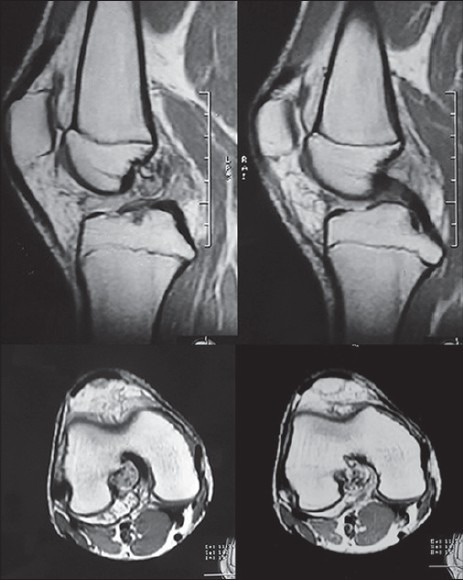
Osteochondromas are usually extra articular and grow away from the joint towards the diaphysis. Intraarticular osteochondromas are very rare and often misdiagnosed. We report a case of 16-year-old boy who presented with pain and clicking sound in the right knee for last 6 months. On examination, click was felt at the terminal flexion of the knee. The lateral radiograph of the right knee showed a radio opaque shadow at the posterior aspect of the distal end of femur, which was further evaluated with an MRI. Arthroscopy showed a hard lesion arising from the roof of the intercondylar notch of femur. It was excised arthroscopically. Histopathology revealed it to be an osteochondroma. Thus, intraarticular osteochondroma of the knee can be considered as a rare cause of pain in young patients.
Keywords: Arthroscopy; intraarticular; knee; osteochondroma.
Conflict of interest statement
Figures


References
-
- Bleshman MH, Levy RM. An unusual location of an osteochondroma. Radiology. 1978;127:456. - PubMed
-
- Takahashi M, Nishihara A, Ohishi T, Shiga K, Yamamoto K, Nagano A. Arthroscopic Resection of an Intraarticular Osteochondroma of the Knee in the Patient with Multiple Osteochondromatosis. Arthroscopy. 2004;20:28–31. - PubMed
-
- Schmoyer S, Ciullo JV. Arthroscopic resection of an osteochondroma of the knee. Arthoscopy. 2001;17:765–7. - PubMed
-
- Dienst M, Schneider G, Pahl S, Ensslin S, Kohn D. Intraarticular osteochondroma of the posterior cavity of the knee. Arch Orthop Trauma Surg. 2002;122:462–5. - PubMed
-
- Siebenrock KA, Ganz R. Osteochondroma of the femoral neck. Clin Orthop Relat Res. 2002;394:211–8. - PubMed
LinkOut - more resources
Full Text Sources
Other Literature Sources