Usefulness of PET/CT for diagnosis of periosteal chondrosarcoma of the femur: A case report
- PMID: 24932240
- PMCID: PMC4049737
- DOI: 10.3892/ol.2014.2010
Usefulness of PET/CT for diagnosis of periosteal chondrosarcoma of the femur: A case report
Abstract
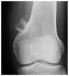
Periosteal chondrosarcoma is an extremely rare low-grade malignant cartilaginous tumor arising from the external bone surface. Diagnosis of periosteal chondrosarcomas may be challenging, since this condition closely resembles periosteal chondromas. It has been reported that positron emission tomography (PET) is useful in distinguishing benign from malignant cartilaginous tumors using a maximum standardized uptake value (SUVmax) cut-off of 2.0 or 2.3. This report presents the case of a 40-year-old female with an 18-month history of a tender mass in the left distal femur. Radiological findings demonstrated periosteal buttressing. Magnetic resonance imaging (MRI) revealed a chondrogenic tumor of 3 cm in size developing from the external bone surface. It was difficult to differentiate periosteal chondrosarcoma from periosteal chondroma on the basis of size and the radiological and MRI findings. PET/computed tomography (CT) revealed abnormal linear uptake with an SUVmax of 2.7, indicating a malignant tumor. A diagnosis of periosteal chondrosarcoma was made, and wide resection was performed. Tumor histology was consistent with grade II chondrosarcoma. PET/CT is thus useful in differentiating periosteal chondrosarcoma from periosteal chondroma.
Keywords: PET; chondrosarcoma; imaging; periosteal tumor.
Figures


Similar articles
-
Periosteal chondroma of the femur: A case report and review of the literature.Oncol Lett. 2015 Apr;9(4):1637-1640. doi: 10.3892/ol.2015.2889. Epub 2015 Jan 23. Oncol Lett. 2015. PMID: 25789014 Free PMC article.
-
Is PET-CT an accurate method for the differential diagnosis between chondroma and chondrosarcoma?Springerplus. 2016 Feb 29;5:236. doi: 10.1186/s40064-016-1782-8. eCollection 2016. Springerplus. 2016. PMID: 27026930 Free PMC article.
-
Periosteal chondroid tumors: radiologic evaluation with pathologic correlation.AJR Am J Roentgenol. 2001 Nov;177(5):1183-8. doi: 10.2214/ajr.177.5.1771183. AJR Am J Roentgenol. 2001. PMID: 11641198
-
Periosteal chondrosarcoma.AJR Am J Roentgenol. 2009 Jan;192(1):W1-6. doi: 10.2214/AJR.08.1159. AJR Am J Roentgenol. 2009. PMID: 19098166 Review.
-
Periosteal chondrosarcoma: a case report and review of the literature.Arch Pathol Lab Med. 1997 Jan;121(1):70-4. Arch Pathol Lab Med. 1997. PMID: 9111097 Review.
References
-
- Schajowicz F. Juxtacortical chondrosarcoma. J Bone Joint Surg Br. 1977;59-B:473–480. - PubMed
-
- Papagelopoulos PJ, Galanis EC, Mavrogenis AF, et al. Survivorship analysis in patients with periosteal chondrosarcoma. Clin Orthop Relat Res. 2006;448:199–207. - PubMed
-
- Kenan S, Abdelwahab IF, Klein MJ, Hermann G, Lewis MM. Lesions of juxtacortical origin (surface lesions of bone) Skeletal Radiol. 1993;22:337–357. - PubMed
-
- Papagelopoulos PJ, Galanis EC, Boscainos PJ, Bond JR, Unni KK, Sim FH. Periosteal chondrosarcoma. Orthopedics. 2002;25:839–842. - PubMed
-
- Robinson P, White LM, Sundaram M, et al. Periosteal chondroid tumors: radiologic evaluation with pathologic correlation. AJR Am J Roentgenol. 2001;177:1183–1188. - PubMed
LinkOut - more resources
Full Text Sources
Other Literature Sources
Molecular Biology Databases
