

Epithelioid angiosarcoma of the spine: A case report of a rare bone tumor
- PMID: 24932310
- PMCID: PMC4049740
- DOI: 10.3892/ol.2014.2055
Epithelioid angiosarcoma of the spine: A case report of a rare bone tumor
Abstract
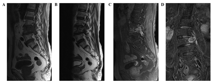
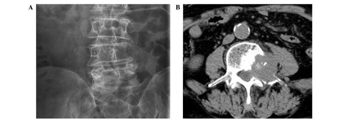
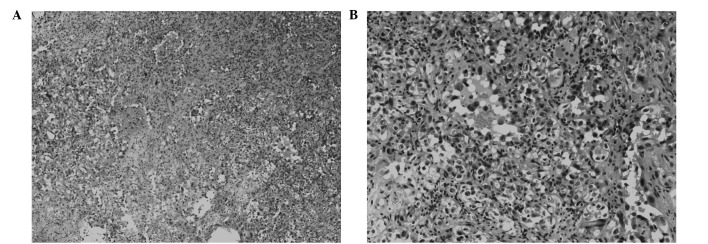
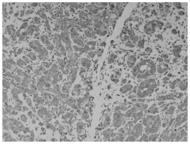
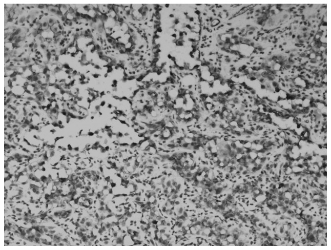
Epithelioid angiosarcoma (EA) is an extremely rare subtype of angiosarcoma, which is characterized by large cells with an epithelioid morphology. EA typically arises in deep soft tissues, including the adrenal gland, skin and thyroid, however, EA rarely arises in the spine. The current study presents a case of osteolytic lesions involving the fourth lumbar (L4) level of the spine. Preoperatively, the patient was misdiagnosed with metastatic carcinoma, however, a radiological examination detected the presence of osteolytic or destructive lesions in the vertebrae, which extended into the pedicles. Histopathological and immunohistochemical evaluations were performed on the tumor tissue obtained from a decompression specimen of the L4 vertebra. A bone lesion composed of sheet-like malignant cells exhibiting atypical epithelioid morphology with vascular formation was observed. The presence of anastomosing vascular channels lined by epithelioid endothelial cells also indicated that focal endothelial differentiation had occurred. In addition, immunohistochemistry assays revealed that the lesion was positive for the endothelial cell markers, CD31, CD34 and vimentin. The tumor was treated with decompression of the L4 vertebra, followed by posterior stabilization. The patient subsequently refused chemotherapy and radiotherapy but completed six months of follow-up. At the time of writing, the tumor remains under control and the patient is asymptomatic. This case highlights the difficulty of diagnosing EA, which requires careful pathological examination and immunophenotype labeling. At present, CD31 is the most sensitive marker for detecting EA.
Keywords: CD31; epithelioid angiosarcoma; factor VIII-related antigen; spine; vertebral tumor.
Figures

Similar articles
-
Multicentric epithelioid angiosarcoma of the spine: a case report of a rare bone tumor.Spine J. 2007 Nov-Dec;7(6):716-9. doi: 10.1016/j.spinee.2006.08.013. Epub 2006 Dec 22. Spine J. 2007. PMID: 17998131
-
Epithelioid angiosarcoma of bone and soft tissue: a report of seven cases with emphasis on morphologic diversity, immunohistochemical features and clinical outcome.Tumori. 2011 Sep-Oct;97(5):585-9. doi: 10.1177/030089161109700508. Tumori. 2011. PMID: 22158488
-
Multicentric epithelioid angiosarcoma of bone.Orthopedics. 2012 Aug 1;35(8):e1293-6. doi: 10.3928/01477447-20120725-39. Orthopedics. 2012. PMID: 22868625
-
Pleural epithelioid angiosarcoma with lymphatic differentiation arisen after radiometabolic therapy for thyroid carcinoma: immunohistochemical findings and review of the literature.Diagn Pathol. 2017 Aug 15;12(1):60. doi: 10.1186/s13000-017-0652-1. Diagn Pathol. 2017. PMID: 28810922 Free PMC article. Review.
-
Two cases of epithelioid angiosarcoma involving the thyroid and a brief review of non-Alpine epithelioid angiosarcoma of the thyroid.Arch Pathol Lab Med. 2003 Feb;127(2):E70-3. doi: 10.5858/2003-127-e70-TCOEAI. Arch Pathol Lab Med. 2003. PMID: 12562256 Review.
Cited by
-
Demonstration of Multiple Metastatic Sites by Positron Emission Tomography/Computed Tomography in a Rare Case of Epithelioid Angiosarcoma of the Scalp.Indian J Nucl Med. 2023 Jan-Mar;38(1):84-86. doi: 10.4103/ijnm.ijnm_92_22. Epub 2023 Feb 24. Indian J Nucl Med. 2023. PMID: 37180187 Free PMC article.
-
Epithelioid angiosarcoma of the cervical spine: A case report.World J Clin Cases. 2025 Jun 16;13(17):101593. doi: 10.12998/wjcc.v13.i17.101593. World J Clin Cases. 2025. PMID: 40524767 Free PMC article.
-
Epithelioid angiosarcoma at chest wall which needs to be carefully distinguished from malignant mesothelioma: report of a rare case.Int J Clin Exp Pathol. 2014 Dec 1;7(12):9056-60. eCollection 2014. Int J Clin Exp Pathol. 2014. PMID: 25674287 Free PMC article.
-
Primary epithelioid angiosarcoma of right hip joint: A case report and literature review.Medicine (Baltimore). 2018 Apr;97(15):e0307. doi: 10.1097/MD.0000000000010307. Medicine (Baltimore). 2018. PMID: 29642158 Free PMC article. Review.
-
Epithelioid angiosarcoma of the ilium: a case report.Int J Clin Exp Pathol. 2014 Dec 1;7(12):9099-103. eCollection 2014. Int J Clin Exp Pathol. 2014. PMID: 25674295 Free PMC article.
References
-
- Errani C, Vanel D, Gambarotti M, et al. Vascular bone tumors: a proposal of a classification based on clinicopathological, radiographic and genetic features. Skeletal Radiol. 2012;41:1495–1507. - PubMed
-
- Hisaoka M, Okamoto S, Aoki T, Yokoyama K, Hashimoto H. Spinal epithelioid hemangioendothelioma with epithelioid angiosarcomatous areas. Skeletal Radiol. 2005;34:745–749. - PubMed
-
- O’Connell JX, Nielsen GP, Rosenberg AE. Epithelioid vascular tumors of bone: a review and proposal of a classification scheme. Adv Anat Pathol. 2001;8:74–82. - PubMed
-
- Skuletić V, Bokun R, Tatomirović Z, Popović L. Epithelioid vascular tumor. Vojnosanit Pregl. 2002;59(Suppl 6):S103–S107. (In Serbian) - PubMed
-
- Balicki D, Buhrmann R, Maclean J, et al. Multicentric epithelioid angiosarcoma of the bone. Pitfalls in clinical and morphological diagnosis. Blood Cells Mol Dis. 1996;22:205–213. - PubMed
LinkOut - more resources
Full Text Sources
Other Literature Sources