Review
. 2014 May 8;4(1):66-73.
eCollection 2014 Jan.

Tendon structure, disease, and imaging

Affiliations
Review

Tendon structure, disease, and imaging

Jeffrey H Weinreb et al. Muscles Ligaments Tendons J. .

Abstract

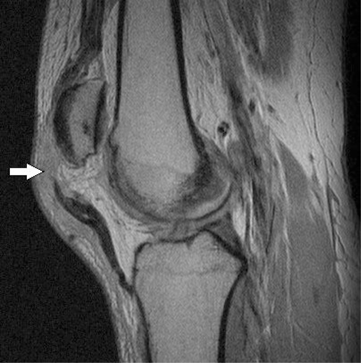
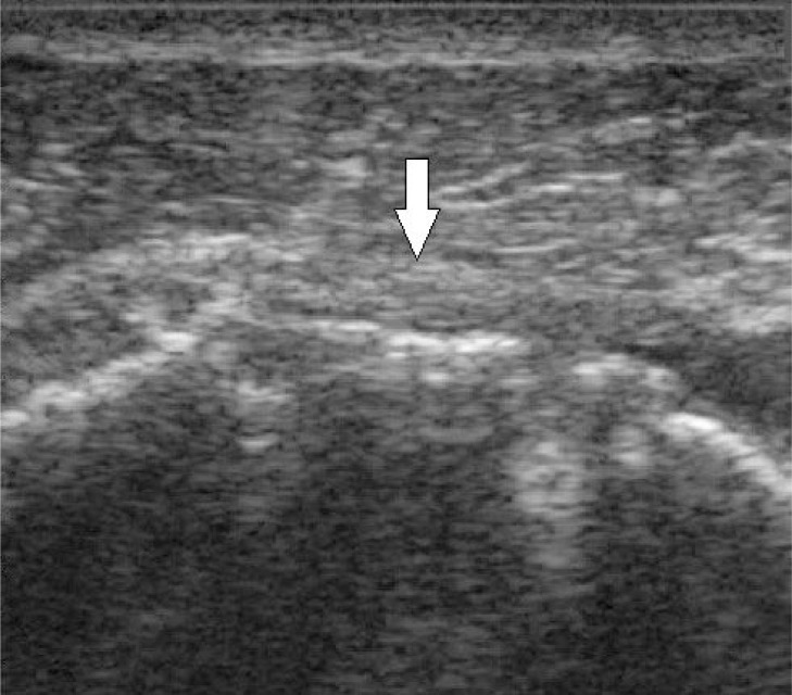
Tendon imaging plays a critical role in evaluating tendon diseases and injuries including mechanical, degenerative, and overuse disease, inflammatory enthesitis, as well as partial and full thickness tears. Ultrasound and magnetic resonance imaging (MRI), each with unique benefits and limitations, are commonly utilized to assist in diagnosing these diseases and conditions. This review delineates important structural properties of tendon and biochemical changes occurring in tendon pathology. This review also examines commonly injured tendons including tendons of the elbow, tendons of the rotator cuff of the shoulder, hip abductor tendons, patellar tendons, and the Achilles tendon to help clinicians better recognize tendon disease. Finally, this paper introduces several emerging imaging techniques including T2 mapping, ultra-short echo time MRI, and sonoelastography as ways in which tendon imaging and evaluation may be improved.

Keywords: Magnetic Resonance Imaging; imaging; tendinopathy; ultrasound.


Figures

Figure 1.
Figure 1.
Axial proton-density weighted MRI of severe insertional bicep tendon partial-thickness tear with tendinopathy.
Figure 2.
Figure 2.
Sagittal proton-density-weighted turbo spin-echo MRI of a full thickness patellar tear with patella alta.
Figure 3.
Figure 3.
Longitudinal ultrasound of an intact supraspinatus tendon one year after successful repair. The arrow demonstrates the parallel echogenic lines representing the fascicles of the tendon.
Figure 4.
Figure 4.
A) Coronal T2 weighted MRI of rotator cuff demonstrating full thickness supraspinatus tendon tear with retraction to the glenoid. B) Coronal T2 weighted MRI of the rotator cuff demonstrating a full thickness supraspinatus tendon tear without retraction.
Figure 5.
Figure 5.
Coronal T2 weighted MRI of supraspinatus tendiosius with reactive subacromial subdeltoid bursitis, intra-substance tear, and hydroxyapatite calcification.
Figure 6.
Figure 6.
Longitudinal ultrasound of a torn supraspinatus tendon three months after surgery. Due to retraction, the echogenic tendon fascicles cannot be visualized. Arrow indicates where the tendon would normally be visualized.
Figure 7.
Figure 7.
Sagittal T2 weight edinsertional Achilles tendinopathy with reactive boney edema pattern (lower arrow) in the calcaneus and retrocalcaneal bursitis (upper arrow).
Figure 8.
Figure 8.
Sagittal T2 weighted MRI of the Achilles tendon demonstrating a full thickness tear with fluid noted by arrow.

References

    1. Birch HL. Tendon matrix composition and turnover in relation to functional requirements. Int J Exp Pathol. 2007;88(4):241–248. - PMC - PubMed
    1. Hodgson R, O’Connor P, Grainger A. Tendon and ligament imaging. Br J Radiol. 2012;85(1016):1157–1172. - PMC - PubMed
    1. Kannus P. Structure of the tendon connective tissue. Scand J Med Sci Sports. 2000;10(6):312–320. - PubMed
    1. Józsa LG, Kannus P. Human tendons: Anatomy, physiology, and pathology. Human Kinetics; Champaign, IL: 1997.
    1. Tresoldi I, Oliva F, Benvenuto M, et al. Tendon’s ultrastructure. Muscles, ligaments and tendons journal. 2013;3(1):2. - PMC - PubMed

LinkOut - more resources