. 2014 Jun 16;9(6):e100292.
doi: 10.1371/journal.pone.0100292. eCollection 2014.

Upper girdle imaging in facioscapulohumeral muscular dystrophy

Affiliations

Upper girdle imaging in facioscapulohumeral muscular dystrophy

Giorgio Tasca et al. PLoS One. .

Abstract

Background: In Facioscapulohumeral muscular dystrophy (FSHD), the upper girdle is early involved and often difficult to assess only relying on physical examination. Our aim was to evaluate the pattern and degree of involvement of upper girdle muscles in FSHD compared with other muscle diseases with scapular girdle impairment.

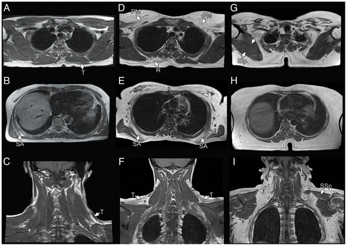
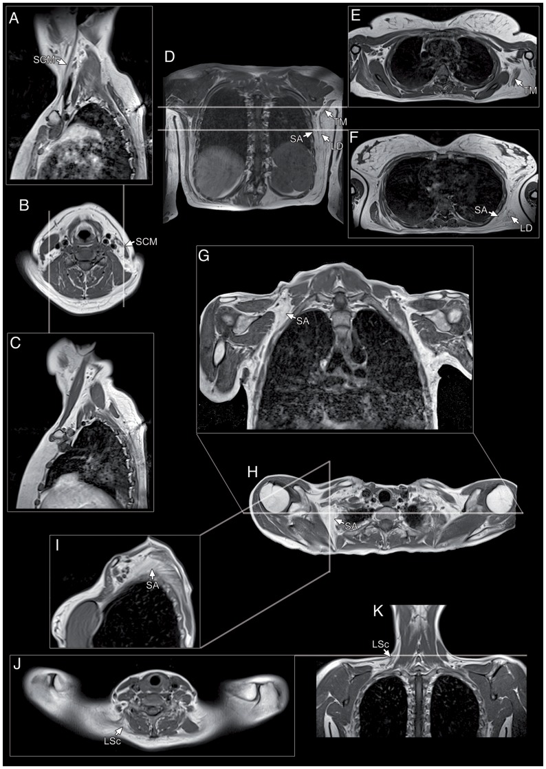
Methods: We propose an MRI protocol evaluating neck and upper girdle muscles. One hundred-eight consecutive symptomatic FSHD patients and 45 patients affected by muscular dystrophies and myopathies with prominent upper girdle involvement underwent this protocol. Acquired scans were retrospectively analyzed.

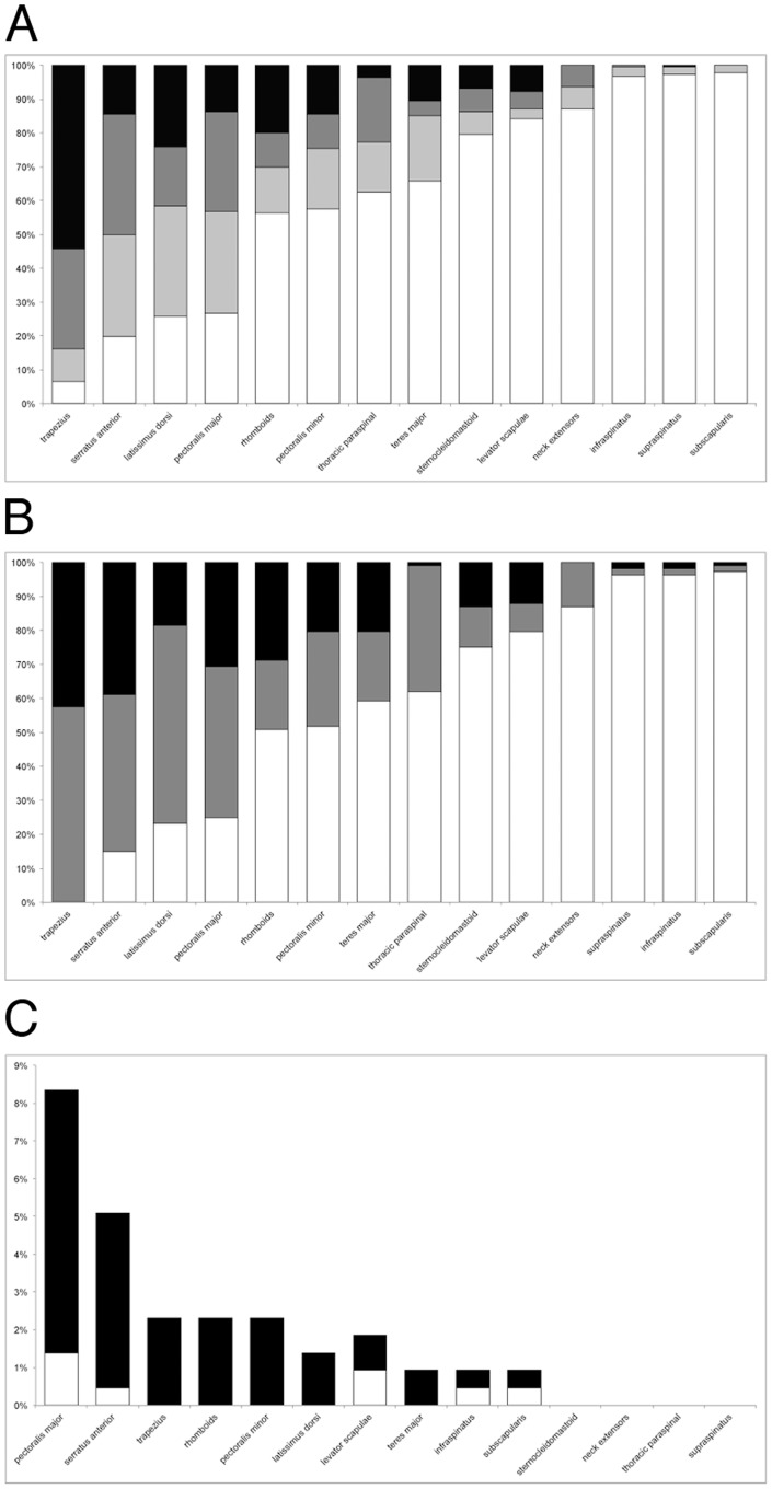
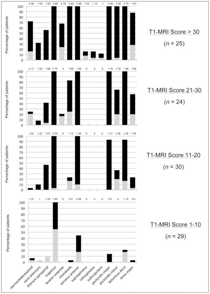
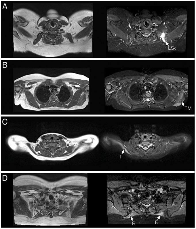
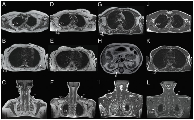
Results: The trapezius (100% of the patients) and serratus anterior (85% of the patients) were the most and earliest affected muscles in FSHD, followed by the latissimus dorsi and pectoralis major, whilst spinati and subscapularis (involved in less than 4% of the patients) were consistently spared even in late disease stages. Asymmetry and hyperintensities on short-tau inversion recovery (STIR) sequences were common features, and STIR hyperintensities could also be found in muscles not showing signs of fatty replacement. The overall involvement appears to be disease-specific in FSHD as it significantly differed from that encountered in the other myopathies.

Conclusions: The detailed knowledge of single muscle involvement provides useful information for correctly evaluating patients' motor function and to set a baseline for natural history studies. Upper girdle imaging can also be used as an additional tool helpful in supporting the diagnosis of FSHD in unclear situations, and may contribute with hints on the currently largely unknown molecular pathogenesis of this disease.


Conflict of interest statement

Competing Interests: The authors of this manuscript have the following competing interests. EM has served on scientific advisory boards for Acceleron Pharma, Prosensa, and PTC Therapeutics; he receives research support from the European Union, Parent Project NL, SMA Europe, and Italian Telethon; he is receiving funding for trials from GlaxoSmithKline; and he has received funding for trials from Trophos and PTC Therapeutics. The other authors have declared that no competing interests exist. The authors also hereby confirm that what is disclosed in the competing interests section does not alter adherence to PLOS ONE policies on sharing data and materials.

Figures

Figure 1
Figure 1. Summary of muscle involvement in FSHD.
(A) Percentage of involvement of the different muscles. White columns: unaffected; light gray columns: affected with score 1; dark gray columns: affected with score 2; black columns: affected with score 3. (B) Frequency of symmetrical and asymmetrical involvement of individual muscles across all the patients. White columns: unaffected; gray columns: affected symmetrically; black columns: affected asymmetrically. (C) Percentage of involvement on STIR sequences of T1-W normal (white) or abnormal (black) muscles.
Figure 2
Figure 2. Graphical representation of the distribution of muscle involvement across different disease severities.
Patients are subdivided into 4 categories of severity according to the T1-MRI score values. The contribution of the progressive involvement of the different muscles to the T1-MRI score is represented as percentage of patients in whom each muscle is affected monolaterally (gray column) or bilaterally (black column) and mean score for each muscle (number on top of the columns).
Figure 3
Figure 3. T1-W MR images in FSHD patients with different disease severity.
In patients with initial disease (A–C) the involvement is generally restricted to the trapezius and serratus anterior muscles, often asymmetrically. In moderately affected patients (D–F), a frequent combination is constituted by bilateral involvement of the trapezius, serratus anterior, pectoralis major and asymmetric rhomboids involvement. In more advanced disease (G–I), other muscles become involved but still with typical complete sparing of the supraspinatus, infraspinatus and subscapularis. T: trapezius; SA: serratus anterior; PM: pectoralis major; R: rhomboids; SSp: supraspinatus; ISp: infraspinatus; SSc: subscapularis.
Figure 4
Figure 4. Identification of asymmetrical muscle involvement in FSHD using different projections.
(A–C) Monolateral atrophy of sternocleidomastoid muscle evident on sagittal and axial sections. Sagittal planes of sectioning used in our protocol were particularly useful to identify and to follow muscles across all their length thus providing a comprehensive assessment on their degree of involvement. (D–F) Coronal section (D) and two axial sections at different levels (E–F) showing asymmetric replacement of the serratus anterior, teres major and latissimus dorsi on one side. (G–I) Involvement of the superior portion of the right serratus anterior can be appreciated in all the three projections used. (J–K) Asymmetric replacement of levator scapulae with sparing of the contralateral on axial and corresponding coronal section. SCM: sternocleidomastoid; SA: serratus anterior; TM: teres major, LD: latissimus dorsi; LSc: levator scapulae.
Figure 5
Figure 5. T1-W images (left) with corresponding STIR images (right).
STIR hyperintensities can be detected in different muscles in absence or with different degree of abnormalities on T1-W images. (A): levator scapulae. (B): teres major. (C) trapezius. (D) rhomboids.
Figure 6
Figure 6. Examples of upper girdle imaging involvement in other myopathies.
LGMD2A (A–C): prominent and symmetric involvement of the serratus anterior, latissimus dorsi and subscapularis muscles with partial sparing of the trapezius. LGMD2B (D–F) major involvement of the supraspinatus, infraspinatus and subscapularis muscles, with relative sparing of the trapezius and serratus anterior. LGMD2L (G–I): isolated involvement of one teres major and thoracic paraspinal muscles, without replacement of trapezius muscles. LGMD1D (J–L): mild involvement of the serratus anterior and subscapularis muscles, again with complete sparing of trapezius. SA: serratus anterior; LD: latissimus dorsi; SSc: subscapularis; T: trapezius; SSp: supraspinatus; ISp: infraspinatus; TM: teres major; TP: thoracic paraspinal.

References

    1. Tasca G, Monforte M, Iannaccone E, Laschena F, Ottaviani P, et al. (2012) Muscle MRI in female carriers of dystrophinopathy. Eur J Neurol 19: 1256–1260. - PubMed
    1. Mercuri E, Pichiecchio A, Allsop J, Messina S, Pane M, et al. (2007) Muscle MRI in inherited neuromuscular disorders: past, present, and future. J Magn Reson Imaging 25: 433–440. - PubMed
    1. Wattjes MP, Kley RA, Fischer D (2010) Neuromuscular imaging in inherited muscle diseases. Eur Radiol 20: 2447–2460. - PMC - PubMed
    1. Padberg GW, van Engelen BG (2009) Facioscapulohumeral muscular dystrophy. Curr Opin Neurol 22: 539–542. - PubMed
    1. Lemmers RJ, van der Vliet PJ, Klooster R, Sacconi S, Camano P, et al. (2010) A unifying genetic model for facioscapulohumeral muscular dystrophy. Science 329: 1650–1653. - PMC - PubMed

Publication types