

Overexpression of piRNA pathway genes in epithelial ovarian cancer
- PMID: 24932571
- PMCID: PMC4059699
- DOI: 10.1371/journal.pone.0099687
Overexpression of piRNA pathway genes in epithelial ovarian cancer
Abstract
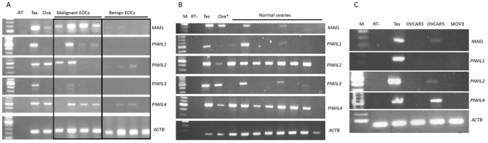
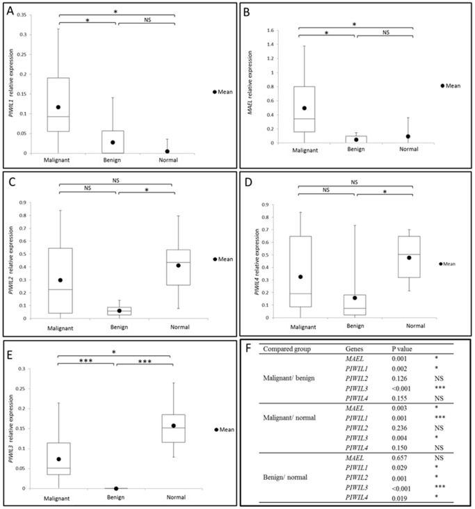
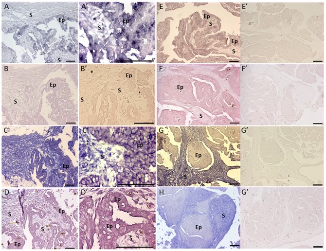
The importance of the Piwi-interacting RNA (piRNA) pathway for germ cell maintenance, genome integrity, DNA methylation and retrotransposon control raises possible roles of this pathway in cancer. Indeed aberrant expression of human PIWI orthologs and Maelstrom has been observed in various cancers. In this study we explored the expression and function of piRNA pathway genes in human ovarian cancer, based on our recent work, which showed widespread expression of piRNA pathway genes in the mammalian. Our work shows that PIWIL1 and MAEL expression is significantly increased in malignant EOC (n = 25) compared to benign tumor tissues (n = 19) and normal ovarian tissue (n = 8). The expression of PIWIL3 is lower in malignant and benign tissues when compared to normal ovary. Sequencing of PIWIL1 transcript revealed that in many tumors deletion of exon 17 leads to the introduction of a premature stop codon in the PIWI domain, likely due to a splicing error. In situ hybridization on tumor sections revealed that L1, PIWIL1, 2 and MAEL are specifically expressed in epithelial cells (cancerous cells) of EOC. Furthermore, PIWIL2 and MAEL are co-expressed in the stromal cells adjacent to tumor cells. Since PIWIL1 and MAEL are up regulated in malignant EOC and expressed in the epithelial cells, we investigated if these two genes affect invasiveness of ovarian cancer cell lines that do not normally express these genes. PIWIL1 and MAEL were transiently over expressed in the ovarian cancer cell line SKOV3, followed by real-time measurements of cell invasiveness. Surprisingly both PIWIL1 and MAEL over expression decreased the invasiveness of SKOV3 cells. Our findings support a growing body of evidence that shows that genes in this pathway are upregulated in cancer. In ovarian cancer we show for the first time that Piwil1 transcript may often be abnormal result in non functional product. In contrast to what has been observed in other cell types, we found that PIWIL1 and MAEL have a repressive effect on cell invasiveness.
Conflict of interest statement
Figures
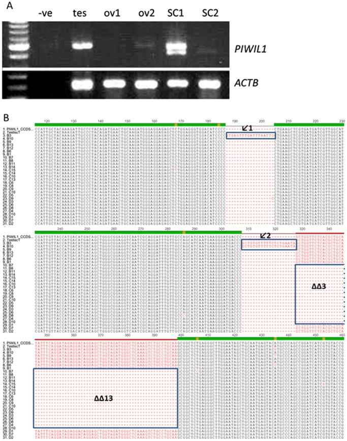
 ). PIWIL1_ccds: published cDNA of PIWIL1 (CCDS9268); Testis C1: testis transcript clone 1; B1–3, B6, B9–B14, C6, C8, C9, C10–C15, D1–D10: clones with PIWIL1 transcripts.
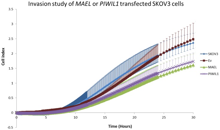
Similar articles
-
Overexpression of PIWI proteins in human stage III epithelial ovarian cancer with lymph node metastasis.Cancer Biomark. 2013;13(5):315-21. doi: 10.3233/CBM-130360. Cancer Biomark. 2013. PMID: 24440970
-
Conservation and expression of PIWI-interacting RNA pathway genes in male and female adult gonad of amniotes.Biol Reprod. 2013 Dec 12;89(6):136. doi: 10.1095/biolreprod.113.111211. Print 2013 Dec. Biol Reprod. 2013. PMID: 24108303
-
PIWIL1 promotes gastric cancer via a piRNA-independent mechanism.Proc Natl Acad Sci U S A. 2020 Sep 8;117(36):22390-22401. doi: 10.1073/pnas.2008724117. Epub 2020 Aug 26. Proc Natl Acad Sci U S A. 2020. PMID: 32848063 Free PMC article.
-
Noncanonical functions of PIWIL1/piRNAs in animal male germ cells and human diseases†.Biol Reprod. 2022 Jul 25;107(1):101-108. doi: 10.1093/biolre/ioac073. Biol Reprod. 2022. PMID: 35403682 Review.
-
A comprehensive review on the role of PIWI-interacting RNA (piRNA) in gynecological cancers.Life Sci. 2024 Nov 15;357:123065. doi: 10.1016/j.lfs.2024.123065. Epub 2024 Sep 17. Life Sci. 2024. PMID: 39299387 Review.
Cited by
-
PIWIL2 promotes progression of non-small cell lung cancer by inducing CDK2 and Cyclin A expression.J Transl Med. 2015 Sep 15;13:301. doi: 10.1186/s12967-015-0666-y. J Transl Med. 2015. PMID: 26373553 Free PMC article.
-
Cancer-testis antigens in ovarian cancer: implication for biomarkers and therapeutic targets.J Ovarian Res. 2019 Jan 4;12(1):1. doi: 10.1186/s13048-018-0475-z. J Ovarian Res. 2019. PMID: 30609934 Free PMC article. Review.
-
The Prognosis Value of PIWIL1 and PIWIL2 Expression in Pancreatic Cancer.J Clin Med. 2019 Aug 22;8(9):1275. doi: 10.3390/jcm8091275. J Clin Med. 2019. PMID: 31443431 Free PMC article.
-
piR-1919609 Is an Ideal Potential Target for Reversing Platinum Resistance in Ovarian Cancer.Technol Cancer Res Treat. 2024 Jan-Dec;23:15330338241249692. doi: 10.1177/15330338241249692. Technol Cancer Res Treat. 2024. PMID: 38706262 Free PMC article.
-
MAEL as a diagnostic marker for the early detection of esophageal squamous cell carcinoma.Diagn Pathol. 2021 Apr 26;16(1):36. doi: 10.1186/s13000-021-01098-z. Diagn Pathol. 2021. PMID: 33902648 Free PMC article.
References
-
- Anttonen M, Ketola I, Parviainen H, Pusa AK, Heikinheimo M (2003) FOG-2 and GATA-4 Are coexpressed in the mouse ovary and can modulate mullerian-inhibiting substance expression. Biol Reprod 68: 1333–1340. - PubMed
-
- Jemal A, Siegel R, Ward E, Hao Y, Xu J, et al. (2008) Cancer statistics, 2008. CA Cancer J Clin 58: 71–96. - PubMed
-
- Ricciardelli C, Oehler MK (2009) Diverse molecular pathways in ovarian cancer and their clinical significance. Maturitas 62: 270–275. - PubMed
-
- Feinberg AP, Vogelstein B (1983) Hypomethylation distinguishes genes of some human cancers from their normal counterparts. Nature 301: 89–92. - PubMed
-
- Lander ES, Linton LM, Birren B, Nusbaum C, Zody MC, et al. (2001) Initial sequencing and analysis of the human genome. Nature 409: 860–921. - PubMed
Publication types
MeSH terms
Substances
LinkOut - more resources
Full Text Sources
Other Literature Sources
Medical
