Review
. 2014 Dec;141(14):1841-55.
doi: 10.1017/S0031182014000626. Epub 2014 Jun 16.

Tools for diagnosis, monitoring and screening of Schistosoma infections utilizing lateral-flow based assays and upconverting phosphor labels

Affiliations
Review

Tools for diagnosis, monitoring and screening of Schistosoma infections utilizing lateral-flow based assays and upconverting phosphor labels

Paul L A M Corstjens et al. Parasitology. 2014 Dec.

Abstract

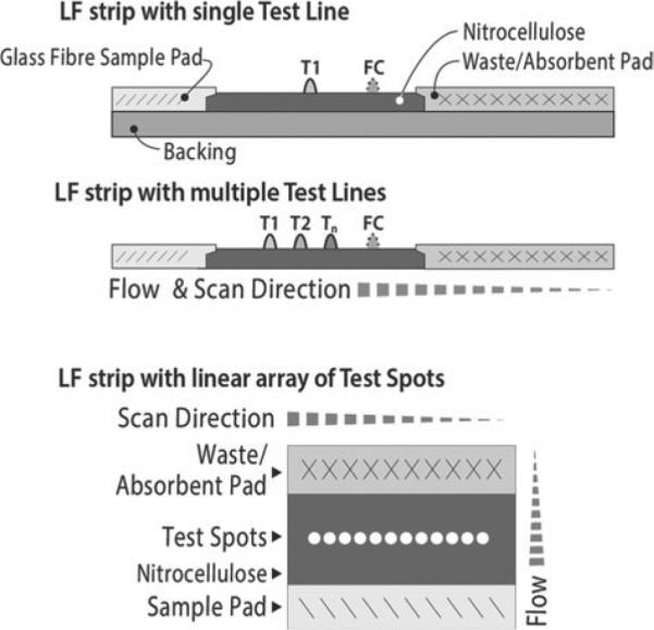
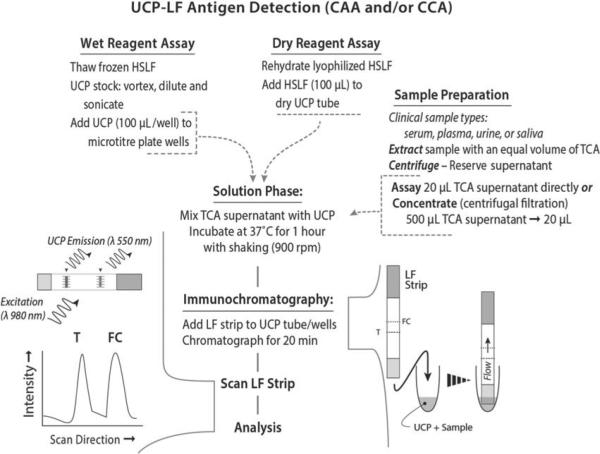
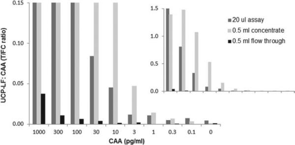
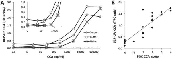
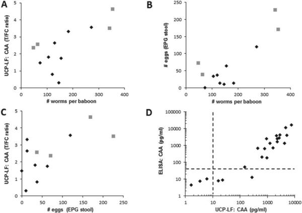
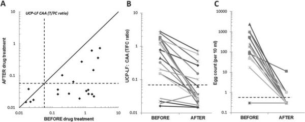
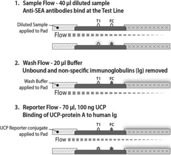
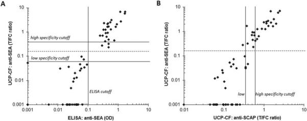
The potential of various quantitative lateral flow (LF) based assays utilizing up-converting phosphor (UCP) reporters for the diagnosis of schistosomiasis is reviewed including recent developments. Active infections are demonstrated by screening for the presence of regurgitated worm antigens (genus specific polysaccharides), whereas anti-Schistosoma antibodies may indicate ongoing as well as past infections. The circulating anodic antigen (CAA) in serum or urine (and potentially also saliva) is identified as the marker that may allow detection of single-worm infections. Quantitation of antigen levels is a reliable method to study effects of drug administration, worm burden and anti-fecundity mechanisms. Moreover, the ratio of CAA and circulating cathodic antigen (CCA) is postulated to facilitate identification of either Schistosoma mansoni or Schistosoma haematobium infections. The UCP-LF assays allow simultaneous detection of multiple targets on a single strip, a valuable feature for antibody detection assays. Although antibody detection in endemic regions is not a useful tool to diagnose active infections, it gains potential when the ratio of different classes of antibody specific for the parasite/disease can be determined. The UCP-LF antibody assay format allows this type of multiplexing, including testing a linear array of up to 20 different targets. Multiple test spots would allow detection of specific antibodies, e.g. against different Schistosoma species or other pathogens as soil-transmitted helminths. Concluding, the different UCP-LF based assays for diagnosis of schistosomiasis provide a collection of tests with relatively low complexity and high sensitivity, covering the full range of diagnostics needed in control programmes for mapping, screening and monitoring.


Figures

Fig. 1
Fig. 1
Different type of lateral flow strips. The upper strip is a side view of a LF strip with a single test (T ) line and a flow-control (FC) line. The T-line is the target-specific (disease/pathogen-specific) capture zone. The FC-line is the control area that captures UCP reporter particles that have flowed past the T-line and indicate that the flow (chromatography) was successful. The LF strip with multiple T-lines can detect a limited number of different targets simultaneously in a single sample; sample and UCP reporter have to pass all capture zones. The lower strip, referred to as TransDot (Malamud et al. 2005; Corstjens et al. 2010), is a top view of a LF strip with a linear array of T-spots; all spots are localized at the same distance from the sample pad and interact with part of the sample and UCP reporter that did not have interaction with the other capture zones. TransDot is well suited for comprehensive multiplexing when it allows the use of a generic UCP conjugate (as UCP particles coated with protein A for antibody detection).
Fig. 2
Fig. 2
UCP-LF antigen detection assay format (CAA and CCA). The UCP-LF antigen test is available as a ‘wet reagent assay’ and a ‘dry reagent assay’. The ‘wet’ format is performed in well-equipped laboratories by qualified staff and has a 3-fold better analytical sensitivity than the ‘dry’ format assay. The ‘dry’ format allows convenient shipping and storage at ambient temperature and is less demanding in equipment and training. Sample preparation is identical for both formats and requires a TCA extraction. The TCA supernatant can be concentrated to increase sensitivity.
Fig. 3
Fig. 3
Concentration devices to improve LLOD. Analysis of an AWA-TCA (containing 3% w/w CAA) standard series and comparison of the 20 μL assay and the 500 μL concentration assay. The standard series was prepared in 1×PBS with 0·5% Tween-20, and extracted with an equal volume of 4% (w/v) TCA; 20 μL of TCA supernatant was analysed with the UCP-LF assay without concentration, and 500 μL TCA-sup was analysed after 25-fold concentration with the Amicon 0·5 mL centrifugal device (10 kDa molecular weight cut-off membrane).
Fig. 4
Fig. 4
Performance of the UCP-CCA assay. In the UCP-CCA assay the anti-CCA antibody #54-4C2 is immobilized at 200 ng per 4 mm on the T line of the LF strip (capture) and covalently coupled to the UCP reporter particle 25 μg per mg UCP reporter (detection). (Panel A) Ratio values determined with the UCP-CCA assay; analysis is of an AWA-TCA (3% w/w CCA) standard series in buffer, urine and serum. (Panel B) The UCP-CCA ratio value of 29 urine reference samples (various origins) compared with the visually recorded semi-quantitative POC-CCA test. The POC-CCA (RMD) scores are semi-quantified as: 0, trace, 1, 2, 3 or 4; 1 indicates the assay threshold below which samples are classified as no response (0) or a trace signal (TS), 1–4 indicate a low, medium, medium-high and high response, respectively.
Fig. 5
Fig. 5
CAA serum levels versus egg and worm count. (Panel A–C) The relation between the number of eggs per gram stool, serum CAA levels expressed as UCP-LF ratio values and the total number of worms counted after perfusion of 8 baboons from previous vaccination studies (diamonds) and 4 baboons from a graded infection experiment (squares, Kariuki and LoVerde, unpublished). (Panel D) Comparison of ELISA CAA concentrations as determined in baboon serum samples from previous vaccinations studies (Kariuki et al. 2004, 2006) vs. UCP-LF CAA concentrations determined during a visit in 2012 to IPR (Nairobi, Kenya) using the same stored serum samples (n = 24). The dotted lines indicate the assay value above which an exponential increase in signal is measured for both assays (van Dam et al. 2013).
Fig. 6
Fig. 6
CAA levels before and after drug treatment. A set of urine samples from 20 individuals before and 2 months after praziquantel treatment (Kahama et al. 1998) were analysed with the 20 μL urine UCP-LF CAA assay (UCAA10, wet reagents). (Panel A) Scatter plot of the UCP-LF ratio values measured in the UCAA10 before and after treatment with praziquantel. Dashed lines indicate the UCP-LF cut-off threshold (0·0571) determined for this particular set of samples. The solid line indicates the ‘no change in CAA concentration’ position; samples with values below this line indicate a decrease of the CAA concentration 2 months after treatment. (Panel B and C) The decrease in CAA concentration (UCP-LF ratio value) and number of eggs (10 mL urine filtrate) showing the respective values ‘before’ and 2 months ‘after’ treatment with praziquantel. Dashed lines indicate cut-off thresholds for the UCP-LF ratio value (0·0571, panel A) and the egg count (arbitrarily set at 0·5 eggs per 10 mL, 1 egg is the minimum number counted).
Fig. 7
Fig. 7
UCP-LF antibody detection format (anti-SEA and anti-SCAP). UCP-LF assay utilizing three sequential flow steps, referred to as consecutive flow (CF). The assay utilizes an Ig-specific label (UCP particles coated with protein A); disease/pathogen specificity is only determined by the T-line (capture zone) on the LF strip. Multiple T-lines can be used as shown in Fig. 1.
Fig. 8
Fig. 8
Performance of two UCP-CF antibody assays. Comparison of the UCP-CF antibody assays with an in-house ELISA to detect anti-SEA antibodies using a set of sera from a previous WHO serum bank, comprised of 28 antibody negatives and 34 antibody positives. (Panel A) The ELISA and UCP-CF performed with SEA. (Panel B) The UCP-CF assays performed with SEA and SCAP. The high and low specificity cut-off thresholds for this experiment are determined according to the protocol described for the antigen assay. The dotted line indicates a threshold for the UCP-CF SEA antibody assay such that the UCP-CF test results match the SEA antibody ELISA. The indicated ELISA cut-off is defined as the average value of the negatives plus 3 s.d.

References

    1. Agnew A, Fulford AJC, de Jonge N, Krijger FW, Rodriguezchacon M, Gutsmann V, Deelder AM. The relationship between worm burden and levels of a circulating antigen (CAA) of 5 species of Schistosoma in mice. Parasitology. 1995;111:67–76. - PubMed
    1. Ayele B, Erko B, Legesse M, Hailu A, Medhin G. Evaluation of circulating cathodic antigen (CCA) strip for diagnosis of urinary schistosomiasis in Hassoba school children, Afar, Ethiopia. Parasitex – Journal de la Societe Francaise de Parasitologie. 2008;15:69–75. - PubMed
    1. Bergquist NR. Schistosomiasis research funding – the TDR contribution. Memorias do Instituto Oswaldo Cruz. 1992;87:153–161. - PubMed
    1. Bergquist R, Johansen MV, Utzinger J. Diagnostic dilemmas in helminthology: what tools to use and when? Trends in Parasitology. 2009;25:151–156. doi: 10.1016/j.pt.2009.01.004. - PubMed
    1. Bergwerff AA, van Dam GJ, Rotmans JP, Deelder AM, Kamerling JP, Vliegenthart JF. The immunologically reactive part of immunopurified circulating anodic antigen from Schistosoma mansoni is a threonine-linked polysaccharide consisting of → 6)-(beta-D GlcpA-(1 → 3))-beta-D-GalpNAc-(1 → repeating units. Journal of Biological Chemistry. 1994;269:31510–31517. - PubMed

Publication types

MeSH terms