. 2014 Dec;29(12):2676-87.
doi: 10.1002/jbmr.2296.

GATA4 is essential for bone mineralization via ERα and TGFβ/BMP pathways

Affiliations

GATA4 is essential for bone mineralization via ERα and TGFβ/BMP pathways

Miriam Güemes et al. J Bone Miner Res. 2014 Dec.

Abstract

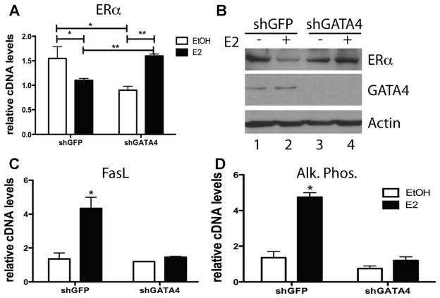
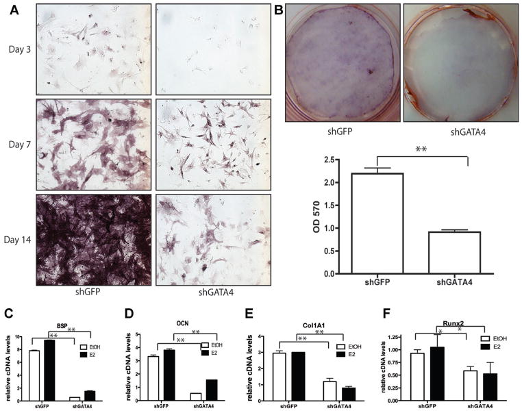
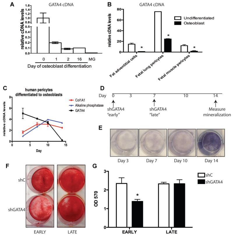
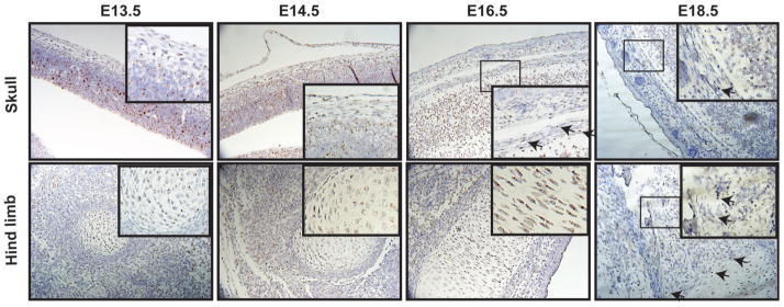
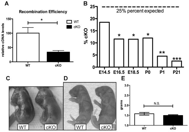
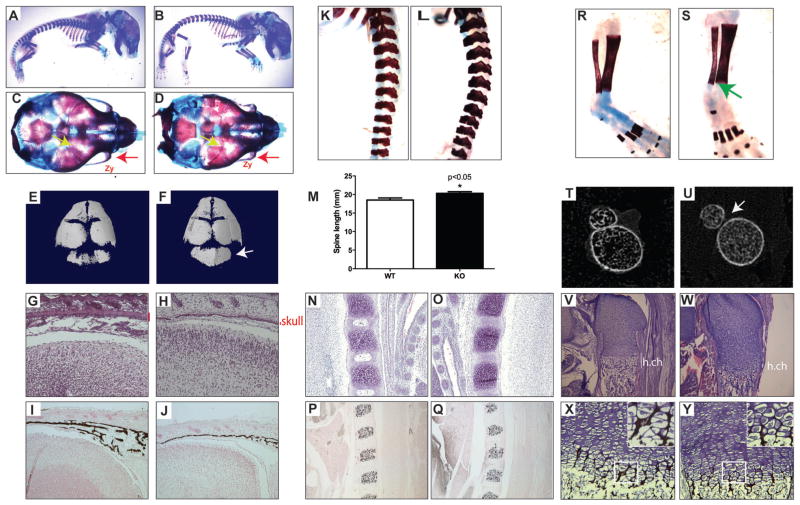
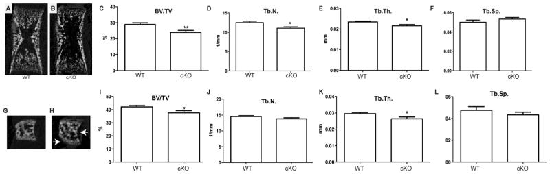
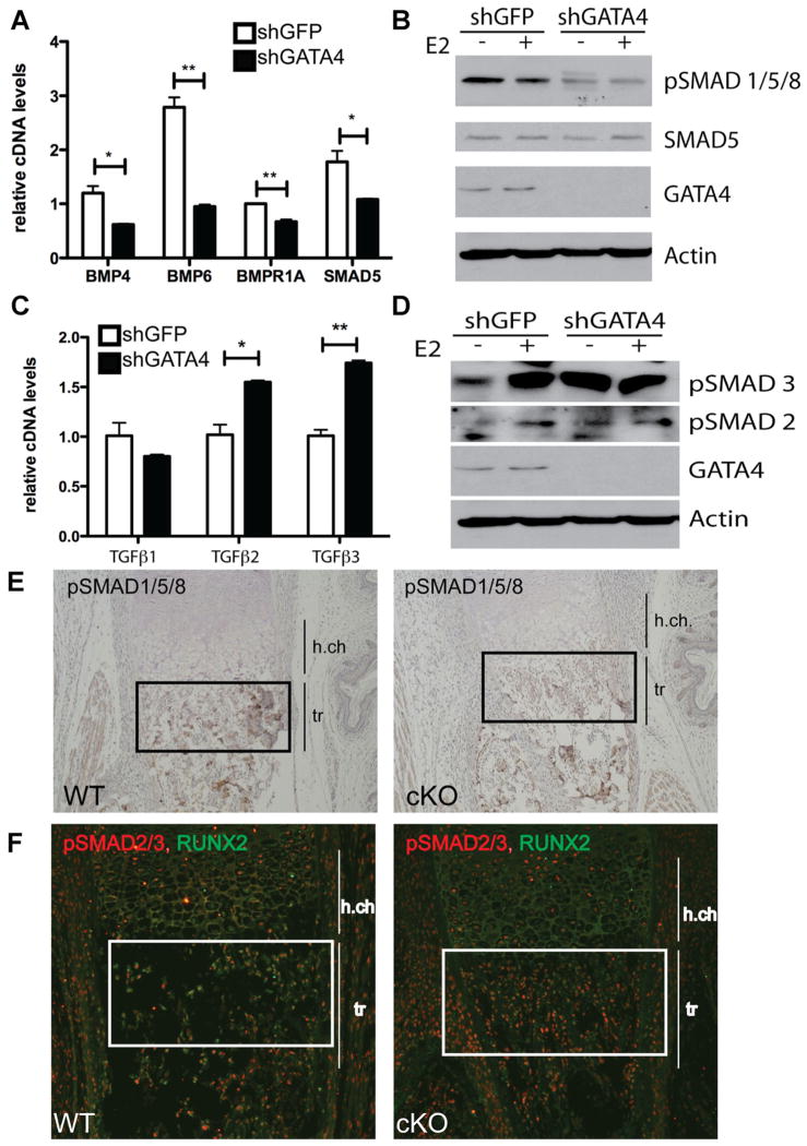
Osteoporosis is a disease characterized by low bone mass, leading to an increased risk of fragility fractures. GATA4 is a zinc-finger transcription factor that is important in several tissues, such as the heart and intestines, and has recently been shown to be a pioneer factor for estrogen receptor alpha (ERα) in osteoblast-like cells. Herein, we demonstrate that GATA4 is necessary for estrogen-mediated transcription and estrogen-independent mineralization in vitro. In vivo deletion of GATA4, driven by Cre-recombinase in osteoblasts, results in perinatal lethality, decreased trabecular bone properties, and abnormal bone development. Microarray analysis revealed GATA4 suppression of TGFβ signaling, necessary for osteoblast progenitor maintenance, and concomitant activation of BMP signaling, necessary for mineralization. Indeed, pSMAD1/5/8 signaling, downstream of BMP signaling, is decreased in the trabecular region of conditional knockout femurs, and pSMAD2/3, downstream of TGFβ signaling, is increased in the same region. Together, these experiments demonstrate the necessity of GATA4 in osteoblasts. Understanding the role of GATA4 to regulate the tissue specificity of estrogen-mediated osteoblast gene regulation and estrogen-independent bone differentiation may help to develop therapies for postmenopausal osteoporosis.

Keywords: ESTROGEN; GATA4; TGF BETA, BMP, OSTEOBLAST.


Conflict of interest statement

Disclosures

All authors state that they have no conflicts of interest.

Figures

Fig. 1
Fig. 1
GATA4 regulates ERα and E2 target genes. (A) Calvarial osteoblasts were infected with lentivirus expressing either shGFP or shGATA4. Cells were then differentiated for 2 wk and RNA was obtained. qPCR was performed for ERα and normalized to actin mRNA. (B) Calvarial osteoblasts were infected with lentivirus expressing either shGFP or shGATA4. Cells were then differentiated for 2 wk and then treated with 10 nM E2 for 24 hours. Protein was obtained and immunoblots for ERα, GATA4, and actin were performed. (C) RNA was obtained as in A, and qPCR was performed with primers to FasL and normalized to actin mRNA. (D) RNA was obtained as in A, and qPCR was performed with primers to alkaline phosphatase and normalized to actin mRNA. *p < 0.05; **p < 0.001. Alk. Phos. = alkaline phosphatase.
Fig. 2
Fig. 2
GATA4 regulates differentiation and mineralization in vitro. (A) Primary calvarial osteoblasts were infected with lentivirus expressing an shRNA directed to GFP (shGFP) or to Gata4 (shGATA4). The following day the cells were placed in mineralization media and allowed to differentiate for 3, 7, or 14 d. Cells were then fixed and assayed for alkaline phosphatase. (B) Primary calvarial osteoblasts were infected with lentivirus expressing an shRNA directed to GFP (shGFP) or to Gata4 (shGATA4). The following day the cells were placed in mineralization media and allowed to differentiate for 2 wk. Cells were then fixed and stained using Alizarin Red. Alizarin Red was eluted and the mineral content was measured at an OD of 570 nm. Silencing was performed in three wells and the average OD is displayed. (CF) Calvarial osteoblasts were infected with lentivirus expressing either shGFP or shGATA4. Cells were then differentiated for 2 wk and RNA was obtained. qPCR was performed for the indicated genes and normalized to actin mRNA. *p < 0.05; **p < 0.001. BSP = bone sialoprotein; OD = optical density; OCN = osteocalcin; Col1A1 = type I collagen A1; Runx, Runt-related transcription factor.
Fig. 3
Fig. 3
GATA4 regulates bone mineralization early in the differentiation process. (A) Bone marrow stromal cells were differentiated for 0, 1, 2, or 16 d. RNA was obtained and qPCR was performed for Gata4 and normalized to actin mRNA. MG RNA was also obtained for comparison. (B) Fat adventitial cells, fetal lung pericytes, and fetal muscle pericytes were left undifferentiated or differentiated to mineralizing osteoblasts. RNA was obtained and qPCR was performed for Gata4 and normalized to actin mRNA. (C) Human fetal bone marrow CD146+ cells (pericytes) were cultured in osteoblast differentiation media for 3, 7, 10, and 14 d. RNA was obtained and qPCR was performed for Gata4, Col1A1, and alkaline phosphatase cDNA and normalized to actin mRNA. (D) Design of experiment for parts EG. (E) Human fetal bone marrow CD146+ cells (pericytes) were cultured in osteoblast differentiation media for 3, 7, 10, and 14 d. At the indicated times cells were fixed and assayed for alkaline phosphatase. (F) Pericytes were infected with lentivirus expressing shC or shGATA4 either “early” at day 0 or “late” at day 7. All cells were placed in mineralization media at day 1 and allowed to differentiate for 2 wk. Cells were then fixed and stained using Alizarin Red. (G) Alizarin Red from part F was eluted and the mineral content was measured at an OD of 570 nm. Silencing was performed in three wells and the average OD is displayed. *p < 0.05. OD = optical density; Col1A1 = type I collagen A1; MG = mammary gland.
Fig. 4
Fig. 4
In situ analysis of Gata4 expression. Sagittal sections of E13.5, E14.5, E16.5, and E18.5 wild-type FVB embryos were probed for Gata4 expression (brown) and counterstained with hematoxylin (blue). Arrows indicate Gata4 expression in bone.
Fig. 5
Fig. 5
GATA4 cKO mice are not born at expected Mendelian ratios. (A) The recombination efficiency of Cre-mediated excision was measured by qPCR from cDNA from differentiated WT and cKO calvarial organ cultures. (B) Tail DNA was obtained from mice at E14.5, E16.5, E18.5, P0, P1, and P21. Genotyping was performed for the presence of Floxed Gata4 and for Cre recombinase. The percent of mice that were GATA4 Fl/Fl/Cre+ (% cKO) is graphed. *p < 0.05; **p < 0.001; ***p < 0.0001. (C) Photograph of representative WT and cKO mice at E18.5. (D) Photograph of representative WT and cKO mice at P1. (E) Weight, in grams, of newborn WT and cKO mice. cKO = conditional knockout; N.S. = not significant.
Fig. 6
Fig. 6
GATA4 cKO mice have skeletal defects. (AD) Skeletal preparations of E18.5 WT (A, C) and cKO (B, D) mice. (A, B) Whole body. (C, D) Superior view of skull. Red arrow indicates decreased zygomatic bone size in cKO mice. Yellow arrows highlight a suture defect in the cKO skull. (E, F) μCT images of WT (E) and cKO (F) skulls. Arrow points to occipital bone. (G, H) H&E staining of sagittal sections of WT (G) and cKO (H) heads. Red line indicates thickness of skull bone. (I, J) Von Kossa staining of sagittal sections of WT (I) and cKO (J) heads. (K, L) Skeletal preparations of spine from WT (K) and cKO (L). (M) Total spine length of n = 7 mice. *p < 0.05. (N, O) H&E staining of sagittal sections of WT (N) and cKO (O) vertebrae. (P, O) Von Kossa staining of sagittal sections of WT (P) and cKO (Q) vertebrae. (R, S) Skeletal preparations of tibia and fibula from WT (R) and cKO (S). Green arrow indicates lack of fusion of tibia and fibula. (T, U) μCT images of WT (T) and ckO (U) tibia and fibula at their closest distance. (V, W) H&E staining of sagittal sections of WT (V) and cKO (W) femurs. (X, Y) Von Kossa staining of sagittal sections of WT (X) and cKO (Y) femurs. Zy = zygomatic bone; h.ch = hypertrophic chondrocytes. KO = knockout; cKO = conditional knockout.
Fig. 7
Fig. 7
Comparison of trabecular bone structure in P0 WT and cKO mice assessed by μCT. (A, B) Representative μCT images of WT and cKO femurs. (C) BV/TV, (D) Tb.N, and (E) Tb.Th were decreased in femurs of cKO mice. (F) There was no statistical increase in Tb.Sp in cKO mice. (G, H) Representative μCT images of WT and cKO L5 vertebrae. Arrows indicate defects in shape. (I) BV/TV, (J) Tb.N, (K) Tb.Th, and (L) Tb.Sp of L5 vertebrae in WT and cKO L5 vertebrae. n = 9; *p < 0.05. cKO = conditional knockout; BV/TV = bone volume/total volume; Tb.N = trabecular number; Tb.Th = trabecular thickness; Tb.Sp = trabecular spacing.
Fig. 8
Fig. 8
GATA4 regulates the TGFβ and BMP pathways in osteoblasts. (A) Calvarial osteoblasts were infected with lentivirus expressing either shGFP or shGATA4. Cells were then differentiated for 2 wk and RNA was obtained. qPCR was performed for the indicated genes and normalized to actin mRNA. (B) Calvarial osteoblasts were infected with lentivirus expressing either shGFP or shGATA4. Cells were then differentiated for 2 wk and then treated with E2 for 24 hours. Protein was obtained and immunoblots for pSMAD1/5/8, SMAD5, GATA4, and actin were performed. (C) RNA was obtained as in A, and qPCR was performed for the indicated genes and normalized to actin mRNA. (D) Protein was obtained as in B, and immunoblots for pSMAD3, pSMAD2, GATA4, and actin were performed. (E) IHC with an antibody to pSMAD1/5/8 was performed on femurs from P0 WT (Fl/Fl) and cKO (Fl/Fl; Cre+) mice. (F) Immunofluorescence with an antibody to pSMAD1/5/8 was performed on femurs from P0 WT (Fl/Fl) and cKO (Fl/Fl; Cre+) mice. *p < 0.05; **p < 0.001. cKO = conditional knockout; h.ch = hypertrophic chondrocytes; tr = trabecular bone; IHC = immunohistochemistry.

References

    1. United States Bone and Joint Decade: The Burden of Musculoskeletal Diseases in the United States. Rosemont, IL: American Academy of Orthopaedic Surgeons; 2008. Available from: http://www.bone-andjointburden.org/chapter_downloads/index.htm.
    1. Kousteni S, Bellido T, Plotkin LI, et al. Nongenotropic, sex-nonspecific signaling through the estrogen or androgen receptors: dissociation from transcriptional activity. Cell. 2001;104(5):719–30. - PubMed
    1. Krum SA, Miranda-Carboni GA, Hauschka PV, et al. Estrogen protects bone by inducing Fas ligand in osteoblasts to regulate osteoclast survival. EMBO J. 2008;27(3):535–45. - PMC - PubMed
    1. Krum SA. Direct transcriptional targets of sex steroid hormones in bone. J Cell Biochem. 2011;112(2):401–8. - PMC - PubMed
    1. Molkentin JD. The zinc finger-containing transcription factors GATA-4, -5, and -6. Ubiquitously expressed regulators of tissue-specific gene expression. J Biol Chem. 2000;275(50):38949–52. - PubMed

Publication types

MeSH terms