Review
. 2014 Aug;57(8):359-68.
doi: 10.1016/j.ejmg.2014.06.002. Epub 2014 Jun 13.

Infantile hydrocephalus: a review of epidemiology, classification and causes

Affiliations
Review

Infantile hydrocephalus: a review of epidemiology, classification and causes

Hannah M Tully et al. Eur J Med Genet. 2014 Aug.

Abstract

Hydrocephalus is a common but complex condition caused by physical or functional obstruction of CSF flow that leads to progressive ventricular dilatation. Though hydrocephalus was recently estimated to affect 1.1 in 1000 infants, there have been few systematic assessments of the causes of hydrocephalus in this age group, which makes it a challenging condition to approach as a scientist or as a clinician. Here, we review contemporary literature on the epidemiology, classification and pathogenesis of infantile hydrocephalus. We describe the major environmental and genetic causes of hydrocephalus, with the goal of providing a framework to assess infants with hydrocephalus and guide future research.

Keywords: Aqueductal stenosis; Genetics; Hydrocephalus; Intraventricular hemorrhage; L1CAM.


Figures

Figure 1
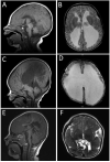
Figure 1. Hydrocephalus without major additional physical findings
A-B: HSAS in a 5-year-old girl with a mutation in L1CAM, for which she was tested only after a mutation was found in her affected younger brother. A: Sagittal T1 demonstrates aqueductal obstruction and stretched, dysplastic or disrupted corpus callosum. B: Axial T2 showing markedly and reduced white matter, particular in the posterior horns. C-D: HSAS in a newborn boy (brother of patient seen in A-B) caused by a mutation in L1CAM. Note similar, though more severe findings when compared to his older sister. C: Sagittal T1 demonstrates complete aqueductal obstruction and stretched corpus callosum. D: Axial T2 showing severely dilated lateral ventricles. E-F: Muscle-eye-brain disease. This girl had been given a diagnosis of aqueductal stenosis at birth. Radiographic features of Muscle-Eye-Brain disease were recognized only after she underwent a repeat MRI at age 3, after which her CK level was tested and found to be markedly elevated she was subsequently found to have a homozygous mutation in POMGNT1. E: Sagittal T1 demonstrates stretched, disrupted corpus callosum, thick tectum (arrow), kinked brainstem and cerebellar cysts. F: Axial T2 reveals cobblestone cortex and abnormal white matter signal in addition to decompressed lateral ventricles.
Figure 2
Figure 2. Hydrocephalus with major additional physical findings
A-C: 9-year-old girl with oro-facial- digital syndrome, Type 1. A: mid-sagittal T1 showing midline cysts with agenesis of the corpus callosum. B, C: axial and coronal T2s showing multiple midline cysts associated with cortical dysplasia. D-F: 6- month-old boy with Chudley-McCollough syndrome. D: sagittal T1 showing third ventricular cyst exerting mass effect on the brainstem and cerebellum. E, F: axial and coronal T2 showing third ventricular cyst, dilated lateral ventricles (particularly the posterior horns) and frontal polymicrogyria (final figure will have arrows). G-I: Hydrocephalus associated with previously unrecognized multisuture synostosis in a 13-month-old girl who underwent a head CT after a breath-holding spell. She was subsequently discovered to have a mutation in FGFR2. Craniosynostosis had not been recognized clinically. G: Sagittal view revealing dilated aqueduct and distal obstruction (posterior fossa crowding with Chiari I malformation). H, I: axial and coronal T2 showing dilated lateral ventricles and absent septum pellucidum.

References

    1. Raimondi AJ. A unifying theory for the definition and classification of hydrocephalus. Childs Nerv Syst. 1994;10(1):2–12. - PubMed
    1. Schrander-Stumpel C, Fryns JP. Congenital hydrocephalus: nosology and guidelines for clinical approach and genetic counselling. Eur J Pediatr. 1998;157(5):355–62. - PubMed
    1. Rekate HL. The definition and classification of hydrocephalus: a personal recommendation to stimulate debate. Cerebrospinal Fluid Res. 2008;5:2. - PMC - PubMed
    1. Munch TN, et al. Familial aggregation of congenital hydrocephalus in a nationwide cohort. Brain. 2012;135(Pt 8):2409–15. - PubMed
    1. Damkier HH, Brown PD, Praetorius J. Cerebrospinal fluid secretion by the choroid plexus. Physiol Rev. 2013;93(4):1847–92. - PubMed

Publication types