. 2014 Oct 17;3(10):731-6.
doi: 10.1021/sb400192a. Epub 2014 Feb 26.

Control of protein function through optochemical translocation

Affiliations

Control of protein function through optochemical translocation

Hanna Engelke et al. ACS Synth Biol. .

Abstract

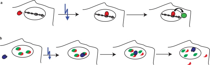
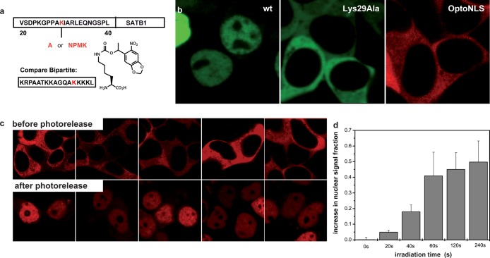
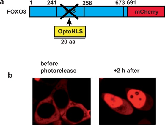
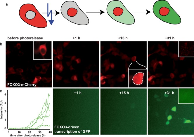
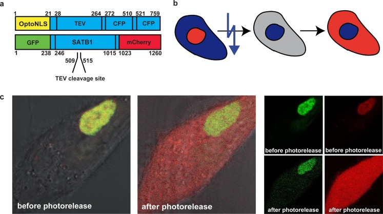
Controlled manipulation of proteins and their function is important in almost all biological disciplines. Here, we demonstrate control of protein activity with light. We present two different applications-light-triggered transcription and light-triggered protease cleavage-both based on the same concept of protein mislocation, followed by optochemically triggered translocation to an active cellular compartment. In our approach, we genetically encode a photocaged lysine into the nuclear localization signal (NLS) of the transcription factor SATB1. This blocks nuclear import of the protein until illumination induces caging group removal and release of the protein into the nucleus. In the first application, prepending this NLS to the transcription factor FOXO3 allows us to optochemically switch on its transcription activity. The second application uses the developed light-activated NLS to control nuclear import of TEV protease and subsequent cleavage of nuclear proteins containing TEV cleavage sites. The small size of the light-controlled NLS (only 20 amino acids) minimizes impact of its insertion on protein function and promises a general approach to a wide range of optochemical applications. Since the light-activated NLS is genetically encoded and optically triggered, it will prove useful to address a variety of problems requiring spatial and temporal control of protein function, for example, in stem-cell, developmental, and cancer biology.

Keywords: nuclear import; optogenetics; photocontrolled TEV-cleavage; photocontrolled transcription; protein control.


Figures

Figure 1
Figure 1
Schematic drawing of the applications: in both cases, light induces nuclear entry of a protein admitting it to its region of activity. (a) A transcription factor (red) is kept in the cytoplasm. Photoactivation releases it to the nucleus where it initiates expression of proteins (green). (b) TEV-protease (blue) is kept in the cytoplasm and enters the nucleus after photoactivation. Proteins in the nucleus (green/red) with a TEV cleavage site will remain intact until the protease enters the nucleus and starts cleavage. Upon cleavage of the protein, its part containing the NLS will remain nuclear (green) whereas the other part (red) will distribute all over the cell.
Figure 2
Figure 2
Photocontrol of SATB1 transcription factor nuclear import. (a) Schematic of light-activated nuclear translocation. The protein of interest is excluded from the nucleus by the photocaged NLS. Upon photorelease of the caging group, the protein enters the nucleus. Lys29 in the SATB1-NLS (aa 20–40) is replaced by alanine or lysine, which is photocaged by the nitropiperonylmethyloxycarbonyl group, blocking nuclear import. For comparison the sequence of the bipartite NLS is shown. (b) SATB1 with wildtype NLS (wt) is located in the nucleus (nuclear signal fraction: 0.950 ± 0.087). eGFP-Lys29Ala-SATB1 and eGFP-OptoNLS-SATB1-mCherry are excluded from the nucleus (nuclear signal fractions: 0.037 ± 0.037 and 0.049 ± 0.026, respectively). (c) Before photolysis, eGFP-OptoNLS-SATB1-mCherry is restricted to the cytoplasm (upper panel). Seven hours after illumination (lasting 120 s) with 350 nm UV light, SATB1 is found predominantly in the nucleus. (d) Dose dependence of the optically gated nuclear import. Difference of nuclear intensity of eGFP-OptoNLS-SATB1-mCherry before illumination and 2 h after a range of UV illumination times.
Figure 3
Figure 3
Photocontrol of transcription factor FOXO3. (a) The construct of OptoNLS-FOXO3-mCherry has the OptoNLS inserted at the site of the native NLS. (b) OptoNLS-FOXO3-mCherry (left) is initially excluded from the nucleus but efficiently enters the nucleus after photorelease (right, 120 s illumination, mean nuclear signal fractions: 0.06 ± 0.03 and 0.64 ± 0.16, respectively).
Figure 4
Figure 4
Photocontrol of FOXO3-mediated transcription. (a) Schematic drawing: A transcription factor (red) is kept in the cytoplasm. Photoactivation releases it to the nucleus where it initiates expression of proteins (green). (b) Nuclear import of OptoNLS-FOXO3-mCherry (red). Before photorelease, OptoNLS-FOXO3-mCherry is excluded from the nucleus. Inset, zoom of single cell. Dashed line denotes nuclear envelope; inset colorscale cropped from 0 to 255 to 0–100 to emphasize absence of detectable leakage into nucleus. After photorelease, OptoNLS-FOXO3-mCherry is actively transported into the nucleus. Inset at t = +15 h: zoom of single cell showing accumulation in nucleus. Dashed line denotes nuclear envelope; inset colorscale cropped as before. (c) Transcriptional activation of a FOXO3-driven GFP reporter (green) upon optically triggered nuclear import of OptoNLS-FOXO3-mCherry. The GFP signal is readily detectable at t > 17 h after photorelease of OptoNLS-FOXO3-mCherry. Each trace in the intensity vs time plot represents one nucleus. The images in parts b and c are the mCherry- and GFP-channel of the same image. The inserts in the last image are a control without UV illumination, where even after 39h no GFP could be detected.
Figure 5
Figure 5
Photocontrol of intranuclear SATB1 cleavage. (a) Photocontrolled delivery of the TEV protease into the nucleus of MCF10A cells and subsequent specific cleavage of a transcription factor. The TEV protease has a prepended OptoNLS and two attached CFPs, to block passive diffusion into the nucleus. A TEV protease site was engineered into the SATB1 transcription factor, which was also flanked by two fluorophores to allow intranuclear cleavage to be visualized. (b) TEV-protease (blue) is kept in the cytoplasm and enters the nucleus after photoactivation. SATB1 in the nucleus (yellow) containing a TEV target site will remain intact until the protease enters the nucleus and starts cleavage. Upon cleavage of the protein, its part containing the NLS will remain nuclear whereas the other part (red) will distribute all over the cell. (c) Fluorescence images: before photorelease of the TEV protease, GFP (green), and mCherry (red) are colocalized and fully contained in the nucleus (left panel), confirming that an intact SATB1 is expressed and localized in the nucleus. After photorelease of TEV, it enters the nucleus and there it cleaves SATB1. The green N-terminal fragment stays in the nucleus since it has a functional NLS; the red C-terminal fragment transitions to the cytoplasm (middle). (right) Images split into single color channels.

References

    1. Barnaszynski L. A.; Wandless T. J. (2006) Conditional control of protein function. Chem. Biol. 13, 11–21. - PubMed
    1. Gardner L.; Deiters A. (2012) Light-controlled synthetic gene circuits. Curr. Opin. Chem. Biol. 16, 292–9. - PMC - PubMed
    1. Riggsbee C. W.; Deiters A. (2010) Recent advances in the photochemical control of protein function. Trends Biotechnol. 28, 468–75. - PMC - PubMed
    1. Yazawa M.; Sadaghiani A. M.; Hsueh B.; Dolmetsch R. E. (2009) Induction of protein–protein interactions in live cells using light. Nat. Biotechnol. 27, 941–945. - PubMed
    1. Polstein L. R.; Gersbach C. A. (2012) Light-inducible spatiotemporal control of gene activation by customizable zinc fincer transcription factors. J. Am. Chem. Soc. 134, 16480–16483. - PMC - PubMed

Publication types

MeSH terms