

Development of antidonor antibody directed toward non-major histocompatibility complex antigens in tolerant animals
- PMID: 24933456
- PMCID: PMC4149823
- DOI: 10.1097/TP.0000000000000249
Development of antidonor antibody directed toward non-major histocompatibility complex antigens in tolerant animals
Abstract
Background: The clinical significance of antibodies directed against antigens other than major histocompatibility complex (MHC) antigens is poorly understood, and there are few large animal models in which such antibodies can be examined. We studied, both retrospectively and prospectively, the development of antibodies to non-MHC antigens in tolerant miniature swine.
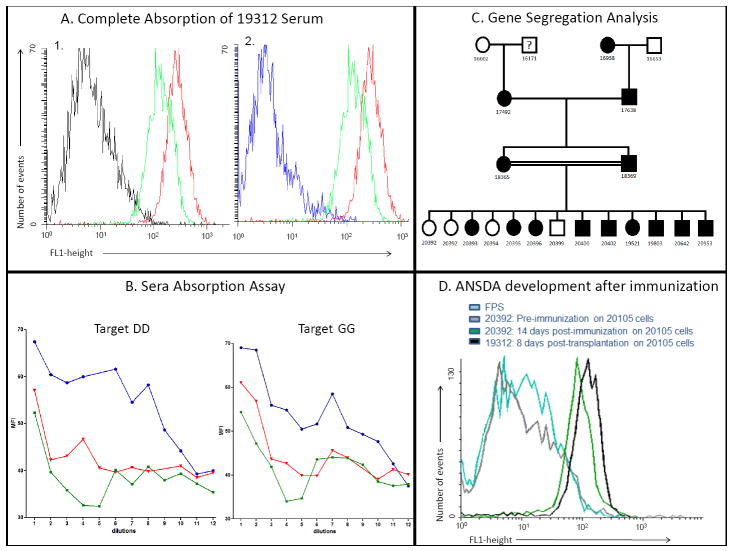
Methods: Our database was assessed for cases of antidonor antibody formation in tolerant animals over the last 20 years. Flow cytometry, absorption assays, and familial analyses for inheritance pattern of the gene(s) potentially responsible for the antibody reactivities were carried out, and an animal determined to be negative for this reactivity was immunized by a skin graft and subcutaneous injections of peripheral blood monocyte cells from an antigen-positive donor.
Results: Sixteen of 469 tolerant animals tested were found to have developed antidonor antibodies. These antibodies were found to be specific for the same, presumably single, non-MHC antigen. Familial analyses indicated that the gene encoding this antigen was expressed in an autosomal-dominant manner in approximately 95% of the herd. In a prospective study, antidonor antibodies with the same specificity as those observed retrospectively were successfully induced in an antigen-negative animal after immunization with peripheral blood monocyte cells.
Conclusion: To our knowledge, this is the first report of the development of antibodies to a highly prevalent, non-MHC antigen present on peripheral blood mononuclear cells and developing in tolerant animals without signs of graft dysfunction. Considering the concern often raised by the appearance of antidonor antibodies in transplant recipients, these data could have important implications for clinical transplantation.
Conflict of interest statement
No conflict of interest for each author
Figures
References
-
- Rosengard BR, Ojikutu CA, Guzzetta PC, et al. Induction of specific tolerance to class I disparate renal allografts in miniature swine with cyclosporine. Transplantation. 1992;54:490–497. - PubMed
-
- Gianello P, Fishbein JM, Sachs DH. Tolerance to primarily vascularized allografts in miniature swine. Immunol Rev. 1993;133:19–44. - PubMed
-
- Rosengard BR, Fishbein JM, Gianello PR, et al. Retransplantation in miniature swine: lack of a requirement for graft adaptation for maintenance of specific renal allograft tolerance. Transplantation. 1994;57:794–799. - PubMed
-
- Tambur AR, Bray RA, Takemoto SK, et al. Flow cytometric detection of HLA-specific antibodies as a predictor of heart allograft rejection. Transplantation. 2000;70:1055–1059. - PubMed
-
- Lee PC, Ozawa M. Reappraisal of HLA antibody analysis and crossmatching in kidney transplantation. Clin Transpl. 2007:219–226. - PubMed
Publication types
MeSH terms
Substances
Grants and funding
LinkOut - more resources
Full Text Sources
Other Literature Sources
Medical
Research Materials
