

Hypermethylation of DDAH2 promoter contributes to the dysfunction of endothelial progenitor cells in coronary artery disease patients
- PMID: 24934151
- PMCID: PMC4069084
- DOI: 10.1186/1479-5876-12-170
Hypermethylation of DDAH2 promoter contributes to the dysfunction of endothelial progenitor cells in coronary artery disease patients
Abstract
Background: Circulating endothelial progenitor cells (EPCs) may be a biomarker for vascular function and cardiovascular risk in patients with coronary artery disease (CAD). Dimethylarginine dimethylaminohydrolase 2 (DDAH2) regulates the function of EPCs. This study aimed to examine whether hypermethylation of DDAH2 promoter contributes to impaired function of EPCs in CAD patients.
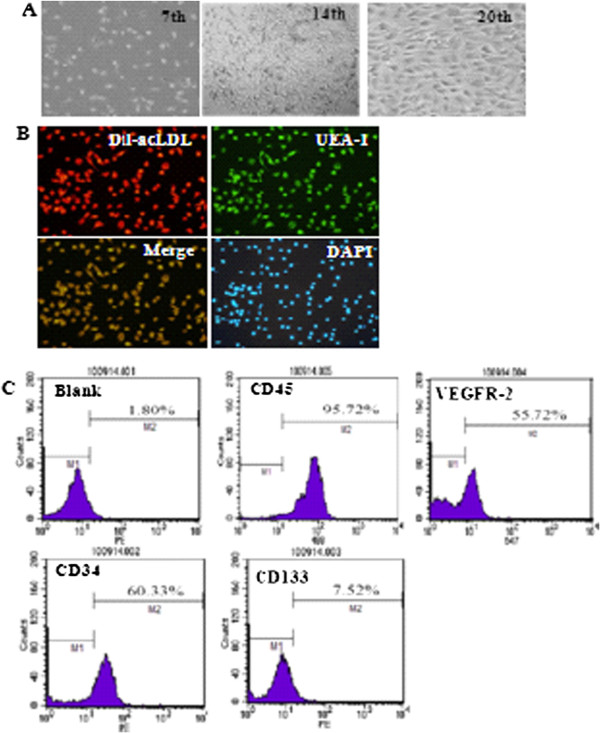
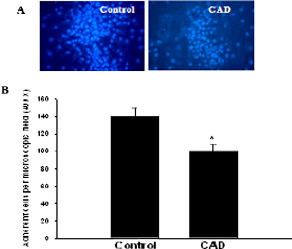
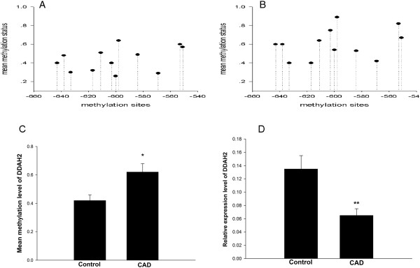
Methods: Peripheral blood mono-nuclear cells from 25 CAD patients and 15 healthy volunteers were collected and differentiated into EPCs. EPCs were tested for their adhesive capability. DDAH2 mRNA expression was analyzed by real-time PCR, and the methylation of DDAH2 promoter was detected by bisulfite genomic sequencing.
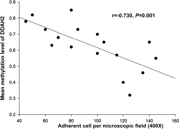
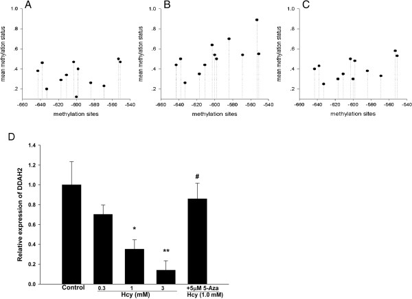
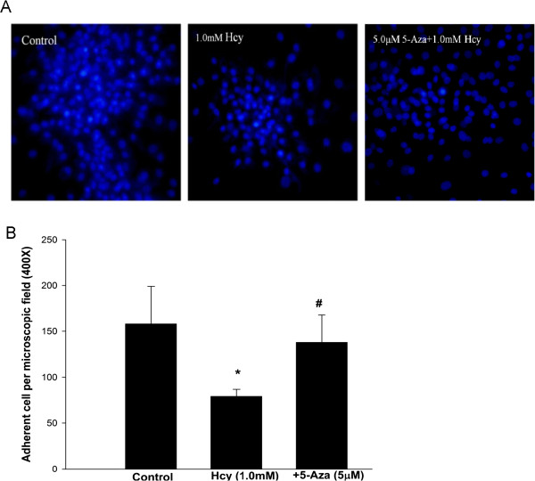
Results: DDAH2 promoter in EPCs from CAD patients was hypermethylated and the methylation level was negatively correlated to DDAH2 mRNA level and adhesion function of EPCs. Homocysteine impaired the adhesion function of EPCs, accompanied by lower DDAH2 expression and higher methylation level of DDAH2 promoter, compared to controls. These effects of homocysteine were reversed by pretreatment with Aza, an inhibitor of DNA methyltransferase.
Conclusion: Hypermethylation in DDAH2 promoter is positively correlated to the dysfunction of EPCs in CAD patients. Homocysteine disrupts EPCs function via inducing the hypermethylation of DDAH2 promoter, suggesting a key role of epigenetic mechanism in the progression of atherosclerosis.
Figures
References
-
- Wassmann S, Werner N, Czech T, Nickenig G. Improvement of endothelial function by systemic transfusion of vascular progenitor cells. Circ Res. 2006;99:e74–e83. - PubMed
-
- Bozdag-Turan I, Turan RG, Paranskaya L, Arsoy NS, Turan CH, Akin I, Kische S, Ortak J, Schneider H, Ludovicy S, Hermann T, D’Ancona G, Durdu S, Akar AR, Ince H, Nienaber CA. Correlation between the functional impairment of bone marrow-derived circulating progenitor cells and the extend of coronary artery disease. J Transl Med. 2012;10:143. - PMC - PubMed
-
- Hill JM, Zalos G, Halcox JP, Schenke WH, Waclawiw MA, Quyyumi AA, Finkel T. Circulating endothelial progenitor cells, vascular function, and cardiovascular risk. N Engl J Med. 2003;348:593–600. - PubMed
-
- Moon JH, Chae MK, Kim KJ, Cha BS, Lee HC, Kim YJ, Lee BW. Decreased endothelial progenitor cells and increased serum glycated albumin are independently correlated with plaque-forming carotid artery atherosclerosis in type 2 diabetes patients without documented ischemic disease. Circ J. 2012;76:2273–2279. - PubMed
Publication types
MeSH terms
Substances
LinkOut - more resources
Full Text Sources
Other Literature Sources
Medical
Miscellaneous
