. 2014 Jul 1;28(13):1461-71.
doi: 10.1101/gad.243832.114. Epub 2014 Jun 16.

Multicilin drives centriole biogenesis via E2f proteins

Affiliations

Multicilin drives centriole biogenesis via E2f proteins

Lina Ma et al. Genes Dev. .

Abstract

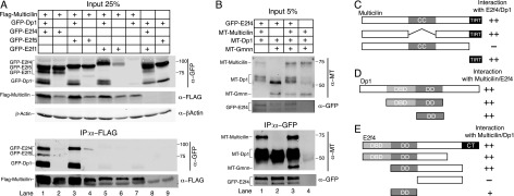
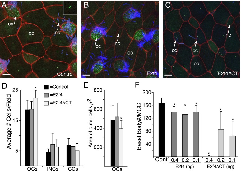
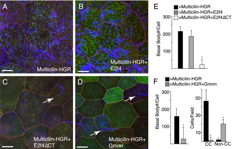
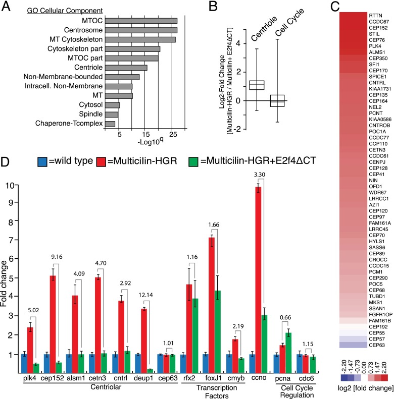
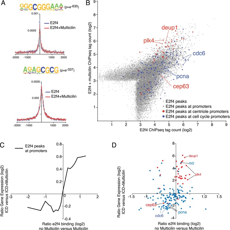
Multiciliate cells employ hundreds of motile cilia to produce fluid flow, which they nucleate and extend by first assembling hundreds of centrioles. In most cells, entry into the cell cycle allows centrioles to undergo a single round of duplication, but in differentiating multiciliate cells, massive centriole assembly occurs in G0 by a process initiated by a small coiled-coil protein, Multicilin. Here we show that Multicilin acts by forming a ternary complex with E2f4 or E2f5 and Dp1 that binds and activates most of the genes required for centriole biogenesis, while other cell cycle genes remain off. This complex also promotes the deuterosome pathway of centriole biogenesis by activating the expression of deup1 but not its paralog, cep63. Finally, we show that this complex is disabled by mutations in human Multicilin that cause a severe congenital mucociliary clearance disorder due to reduced generation of multiple cilia. By coopting the E2f regulation of cell cycle genes, Multicilin drives massive centriole assembly in epithelial progenitors in a manner required for multiciliate cell differentiation.

Keywords: centrioles; e2f4; multiciliate cells.


Figures

Figure 1.
Figure 1.
Multicilin interactions with E2f family members and Gmnn. (A,B) Extracts of stage 12 animal caps were prepared from Xenopus embryos that were injected with the indicated RNAs and subjected to Western analysis with the indicated antibody before (input) and after immunoprecipitation (IP). (C–E) The domain structures of Multicilin (C), Dp1 (D), and E2f4 (E) are diagrammed along with deletion mutants that were tested for their ability to form the EDM complex, summarizing data shown in Supplemental Figure 2.
Figure 2.
Figure 2.
MCC differentiation is blocked in the skin of E2f4ΔCT-expressing embryos. (A–C) Shown are confocal images of embryonic skin (stage 28) of control embryos (A) or embryos injected with RNA encoding E2f4 (B) or E2f4ΔCT (C). Embryos were also injected with RNA encoding mRFP (red) and Hyls1-GFP (green) to mark membranes and basal bodies/centrioles, respectively, and stained for cilia (blue). Cells extending one or two cilia are marked with an arrow in C. Different cell types are identified based on morphology and cilia staining as outer cells (OCs), MCCs, small intercalated cells (INCs), and ciliated cells (CCs). Bar, 10 μm. (D) Representation of different skin cell types in control or E2f4- or E2f4ΔCT-expressing embryos based on 12 fields (98 μm2) from six embryos. (E) Effects on the cell cycle can be indirectly read out by the size of outer cells (Stubbs et al. 2012). E2f4ΔCT significantly (P < 0.05) decreases average cell size based on 24 cells from six embryos, indicating that it weakly promotes cell division. (F) Basal body number in MCCs in control or E2f4- or E2f4ΔCT-expressing embryos. Basal body counts based on 10 to 15 MCCs taken from six embryos. In all graphs, error bars indicate SD, and values significantly different (P < 0.05) from controls based on a two-tailed t-test are marked (asterisks).
Figure 3.
Figure 3.
E2f4ΔCT and Gmnn block Multicilin-induced basal body assembly during MCC differentiation. (A–D) Shown are confocal images of the embryonic skin expressing Multicilin-HGR alone (A) or with E2f4 (B), E2f4ΔCT (C), or Gmnn (D). Multicilin-HGR activity was induced with DEX at stage 12 and embryos fixed at stage 28. Membranes are marked with mRFP (red), basal bodies with Hyls1-GFP (green), and cilia in blue. Arrows denote monociliated cells. Bar, 10 μm. (E) Basal body number induced by ectopic expression of Multicilin-HGR activity in the presence of E2f4 and E2f4ΔCT. (F) Basal body number or the number of ciliated cells per field induced by Multicilin-HGR alone or in the presence of Gmnn. Basal body counts based on 10 to 15 cells taken from six embryos. Error bars indicate SD, and values significantly different (P < 0.05) from controls based on a two-tailed t-test are marked (asterisks)
Figure 4.
Figure 4.
E2f4ΔCT inhibits centriole gene expression during MCC differentiation. (A) RNA-seq analysis of skin progenitors isolated from Xenopus embryos expressing Multicilin-HGR alone or with E2f4ΔCT. Skin progenitors were isolated at stage 10, treated with DEX at stage 11 to induce MCC differentiation, and then extracted for RNA 9 h later. GO terms (P < 0.01) for genes down-regulated by E2f4ΔCT with the highest significance for microtubule (MT)-associated structures, including centrioles. GO term analysis for genes up-regulated by E2f4ΔCT failed to find a significant term. (B) Tukey box plot showing fold changes in response to E2f4ΔCT in the background of Multicilin-HGR-induced differentiation for both centriole components based on the supplemented list generated by Azimzadeh et al. (2012) or for cell cycle genes (Kegg Pathway) (Supplemental Tables 2, 3) (C) Heat map showing log2 fold change in response to E2f4ΔCT in Multicilin-HGR samples. (D) RNA samples were generated as in A but then analyzed in triplicate for the expression of the indicated gene using quantitative RT–PCR. RNA levels are shown after normalization to ubiquitously expressed ornithine decarboxylase (odc) RNA and are set relative to a value of 1 for uninjected controls. Fold change in the presence and absence of E2f4ΔCT is indicated.
Figure 5.
Figure 5.
E2f4-binding sites in skin progenitors and differentiating MCCs. ChIP-seq analysis was carried out on skin progenitors expressing E2f4-GFP in the presence or absence of Multicilin-HGR. Recovered DNA was sequence-aligned to the X. laevis genome, peaks were called, and tags within those peaks were counted. (A) Top two motifs enriched in E2f4 ChIP-seq and the positions of those motifs in immunoprecipitated genomic DNA from both experiments. Motif statistics are from E2f4 + Multicilin; for other conditions, see Supplemental Figure 6. (B) Tag densities for all E2f4 peaks, E2f4 peaks in promoters (±1 kb from the transcription start site), E2f4 peaks in the promoters of centriole genes, and E2f4 peaks in the promoters of cell cycle genes in the presence or absence of Multicilin. Centriole genes are strongly bound by E2f4 and even more so in the presence of Multicilin. (C) Moving average of the ratio of expression of all genes with promoters bound by E2f4 in the presence or absence of Multicilin. Note the general increase in transcription of genes with promoters more strongly bound in the presence of Multicilin. (D) Ratio of expression of centriole and cell cycle genes and binding of E2f4-GFP in the presence or absence of Multicilin.
Figure 6.
Figure 6.
Mutations that cause human RGMC affect EDM complex formation and function. (A) Extracts of skin progenitors (stage 12) isolated from embryos injected with the indicated RNAs were subjected to Western blot analysis prior to (input) or after immunoprecipitation (IP) using the indicated antibodies. (B–E) Shown are confocal images of the skin in embryos expressing Multicilin-R370H and/or E2f4ΔCT-VP16 as indicated. Membranes are marked with mRFP (red), basal bodies are marked with Chibby-GFP (green), and cilia are labeled in blue. (F) The percentage of MCCs in the skin for each RNA injection. Values that differ significantly from the control based on a two-tailed t-test are marked ([*] P < 0.01). (G–K) Shown are confocal images of the skin in embryos expressing activated Notch (ICD) alone or with Multicilin-R370H and/or E2f4ΔCT-VP16 as indicated. Membranes are marked with mRFP (red), basal bodies are marked with Chibby-GFP (green), and cilia are labeled in blue. (L) The percentage of cells that are MCCs in the skin for each RNA injection. Values for Multicilin- and Multicilin-R370H/E2f4ΔCT-VP16-expressing embryos are not significantly different. Data were obtained from 10 fields from five embryos. Error bars indicate SD. Bars, 10 μm.

Comment in

References

    1. Azimzadeh J, Wong ML, Downhour DM, Sanchez Alvarado A, Marshall WF 2012. Centrosome loss in the evolution of planarians. Science 335: 461–463 - PMC - PubMed
    1. Bettencourt-Dias M, Rodrigues-Martins A, Carpenter L, Riparbelli M, Lehmann L, Gatt MK, Carmo N, Balloux F, Callaini G, Glover DM 2005. SAK/PLK4 is required for centriole duplication and flagella development. Curr Biol 15: 2199–2207 - PubMed
    1. Blythe SA, Reid CD, Kessler DS, Klein PS 2009. Chromatin immunoprecipitation in early Xenopus laevis embryos. Dev Dyn 238: 1422–1432 - PMC - PubMed
    1. Boon M, Wallmeier J, Ma L, Loges NT, Jaspers M, Olbrich H, Dougherty G, Werner C, Amirav I, Hevroni A, et al. 2014. Mutations in MCIDAS result in a congenital mucociliary clearance disorder with reduced generation of multiple motile cilia (RGMC). Nat Commun (in press) - PubMed
    1. Carroll SB 2008. Evo-devo and an expanding evolutionary synthesis: a genetic theory of morphological evolution. Cell 134: 25–36 - PubMed

Publication types