. 2014 Jun;6(6):481-95.
doi: 10.18632/aging.100673.

Interaction between ROS dependent DNA damage, mitochondria and p38 MAPK underlies senescence of human adult stem cells

Affiliations

Interaction between ROS dependent DNA damage, mitochondria and p38 MAPK underlies senescence of human adult stem cells

Aleksandra Borodkina et al. Aging (Albany NY). 2014 Jun.

Abstract

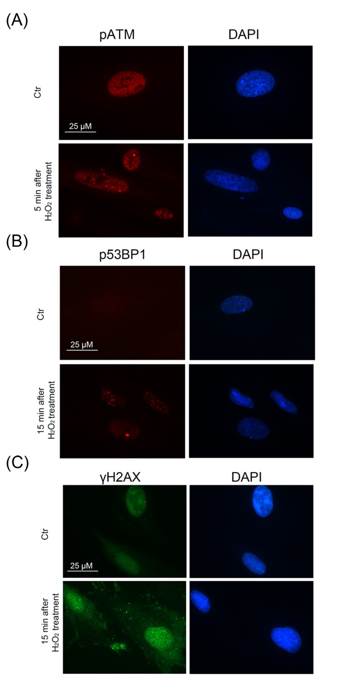
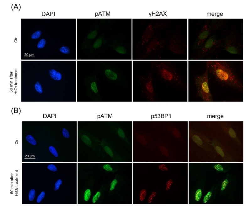
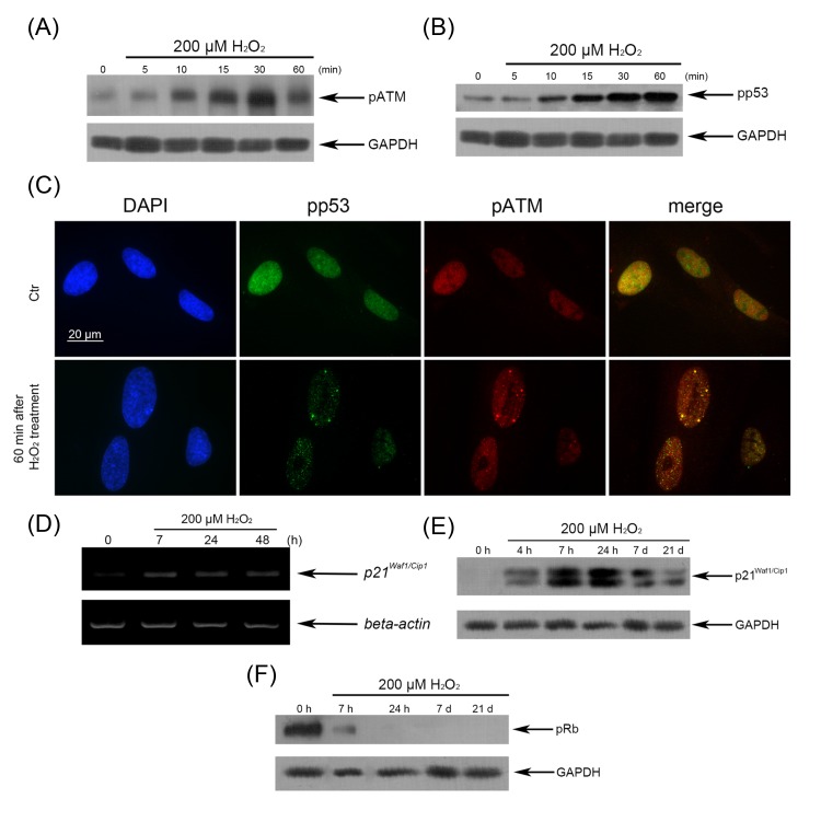
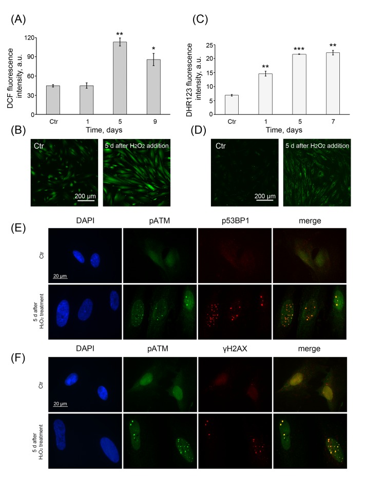
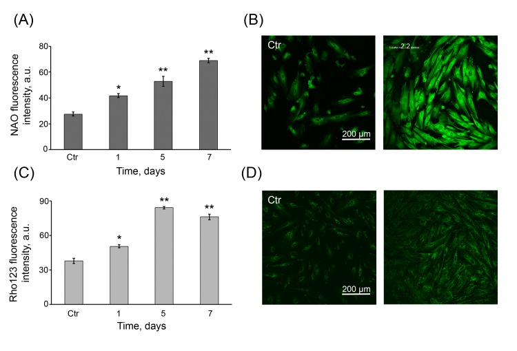
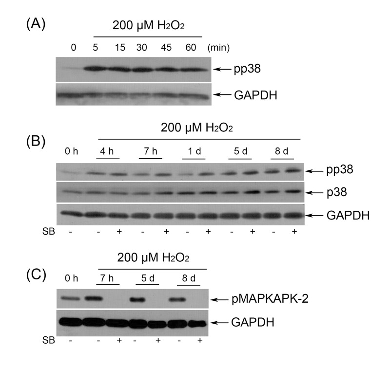
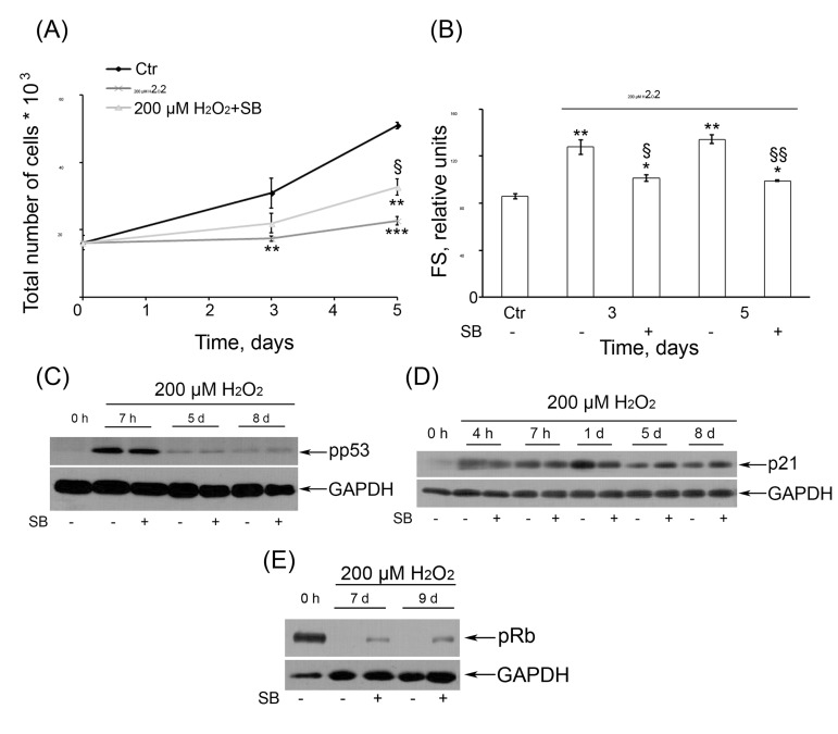
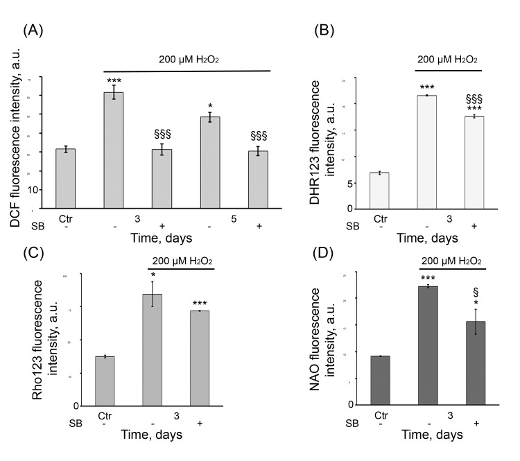
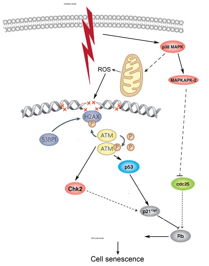
Human endometrium-derived mesenchymal stem cells (hMESCs) enter the premature senescence under sublethal oxidative stress, however underlying mechanism remains unknown. Here, we showed that exogenous H2O2 induces a rapid phosphorylation and co-localization of ATM, H2A.X, 53BP1 leading to DNA damage response (DDR) activation. DDR was accompanied with nuclear translocation of p-p53 followed by up-regulation of p21Waf1 and the permanent hypophosphorylation of pRb. Additionally, the increased p38MAPK/MAPKAPK-2 activation persisted in H2O2-treated cells. We suggest that both p53/p21/pRb and p38MAPK/MAPKAPK-2 pathways are responsible for establishing an irreversible cell cycle arrest that is typical of senescence. The process of further stabilization of senescence required prolonged DDR signaling activation that was provided by the permanent ROS production which in turn was regulated by both p38MAPK and the increased functional mitochondria. To reverse senescence, the pharmacological inhibition of p38MAPK was performed. Cell treatment with SB203580 was sufficient to recover partially senescence phenotype, to block the ROS elevation, to decrease the mitochondrial function, and finally to rescue proliferation. Thus, suppression of the p38MAPK pathway resulted in a partial prevention of H2O2-induced senescence of hMESCs. The current study is the first to reveal the molecular mechanism of the premature senescence of hMESCs in response to oxidative stress.


Conflict of interest statement

Authors declare no conflict of interests.

Figures

Figure 1
Figure 1. The dynamics of H2O2 penetration into hMESCs
(A) Intracellular ROS levels upon application of 200 µM H2O2. Cells were loaded with DCF, each trace represents the response of one cell. Three (N = 3) experiments were performed. (B) Fluorescent images taken at different stages (indicated by dashed lines) of the experiment illustrated in (A). Scale bar is 200 µm and valid for all images. (C) Intracellular ROS levels measured by FACS after staining with the fluorescent probe DCF. Values are means ± SD of three independent experiments. **p<0.005, ***p<0.001 versus control.
Figure 2
Figure 2. A rapid DDR activation in H2O2-treated hMESCs
Immunofluorescence analysis of ATM (A), 53BP1 (B) activation and γH2AX foci formation (C) performed at indicated time points after H2O2 addition. DAPI was used as nuclear stain (blue). Representative photomicrographs of the staining are made at original magnification X100. Scale bar is 25 µm.
Figure 3
Figure 3. Formation of DNA damage foci containing pATM, γH2AX, p53BP1 in H2O2-treated hMESCs
Immunofluorescent analysis with the use of specific antibodies against pATM, γH2AX, p53BP1 revealed co-localization of pATM with either γH2AX (A) or p53BP1 (B) in 60 min after beginning of H2O2 treatment. DAPI was used as nuclear stain (blue). Images are taken at magnification X100. Scale bar is 20 µm and valid for all images.
Figure 4
Figure 4. H2O2-induced activation of the p53/p21/pRb pathway in hMESCs
(A) H2O2-induced ATM phosphorylation tested by Western blotting. (B) Phosphorylation status of p53 in both untreated and H2O2-treated cells examined by Western blotting. (C) Nuclear accumulation of pp53 and co-localization pp53 with pATM revealed by immunofluorescence. Original magnification, X100. Scale bar, 20 µm. (D) The expression levels of p21 mRNA. (E) The expression levels of p21 protein. (F) Abrogation of Rb phosphorylation in H2O2-treated hMESCs examined by Western blotting. GAPDH and β-actin were used as loading controls. Representative results of three independent experiments are shown.
Figure 5
Figure 5. Permanent ROS generation and prolonged DDR activation
(A) Intracellular ROS levels and (C) cellular peroxide levels detected at indicated time points by FACS analysis after staining with either DCF or DHR123, respectively. (M ± SD, N = 3, *p<0.05, **p<0.005, ***p<0.001, versus control). Representative DCF (B) or DHR123 (D) fluorescent images of control (Ctr) and treated hMESCs. Scale bar is 200 µm and valid for all images. (E) Co-localization of pATM with either p53BP1 or (F) γH2AX in 5 days after H2O2 treatment. DAPI was used as nuclear stain (blue). Representative photomicrographs of the staining are shown. Images are taken at magnification X100. Scale bar, 20 µm.
Figure 6
Figure 6. Persistent increase in the number of functional mitochondria in H2O2-treated hMESCs
(A) FACS analysis of mitochondrial mass in control (Ctr) and H2O2-treated hMESCs stained with NAO. Values are means ± SD of three independent experiments (*p<0.05, **p<0.005, versus control). (B) Representative NAO confocal fluorescence images of control (Ctr) and treated hMESCs. (C) Rho123 fluorescence in hMESCs at the indicated time points after H2O2 treatment as measured by FACS. (M ± SD, N = 3, *p<0.05, **p<0.005, versus control). (D) Rho123 fluorescent images of control (Ctr) and H2O2-treated hMESCs. Micrographs are representative for three experiments. Scale bar is 200 µm and valid for all images.
Figure 7
Figure 7
(A) Immunoblot analysis of p38 phosphorylation during 1 h H2O2 treatment. (B) SB had no effect on long-term p38 phosphorylation as detected by Western blot. (C) Selective inhibition of p38 activity abolished phosphorylation of MAPKAPK-2 during the whole observation period. Representative results of three independent experiments are shown. GAPDH was used as loading control.
Figure 8
Figure 8
(A) SB rescued the cell proliferation of H2O2-treated hMESCs. Cell number was determined daily by FACS (M ± SD, N = 3, **p<0.005, ***p<0.001, versus control, §p<0.05, versus H2O2-treated cells). (B) SB partially prevented H2O2-induced increase of cell size. Forward scatter (FS) reflects the average cell size. M ± SD, N = 3, *p<0.05, **p<0.005, versus control, §p<0.05, §§<0.005, versus H2O2-treated cells. (C) Inhibition of p38 activity had no effect on long-term activation of pp53 or (D) p21. (E) An impact of inhibition of p38 kinase activity on pRb phosphorylation status.
Figure 9
Figure 9. SB diminished ROS production and mitochondrial function in senescent hMESCs
(A) DCF, (B) DHR123, (C) Rho123, and (D) NAO fluorescence in control (Ctr), H2O2-treated and (SB+H2O2)-treated hMESCs at indicated time points as measured by flow cytometry (M ± SD, N = 3, *p<0.05, ***p<0.001, versus control, §p<0.05, §§§<0.001, versus H2O2-treated cells).
Figure 10
Figure 10. A pathway interaction scheme displaying the proposed molecular mechanism of premature sense-cence of hMESCs under oxidative stress

References

    1. Campisi J. Senescent cells, tumor suppression, and organismal aging: good citizens, bad neighbours. Cell. 2005;120:513–522. - PubMed
    1. Minamino T, Komuro I. Vascular cell senescence: contribution to atherosclerosis. Circ Res. 2007;100:15–26. - PubMed
    1. Sone H, Kagawa Y. Pancreatic beta cell senescence contributes to the pathogenesis of type 2 diabetes in high-fat diet-induced diabetic mice. Diabetologia. 2005;48:58–67. - PubMed
    1. Adams PD, Sedivy JM. Cellular Senescence and Tumor Suppression. Springer New York Dordrecht Heidelberg London. 2010. p. 272.
    1. Sethe S, Scutt A, Stolzing A. Aging of mesenchymal stem cells. Ageing Res Rev. 2006;5:91–116. - PubMed

Publication types

MeSH terms

Substances