

Quantification of skeletal asymmetries in normal adolescents: cone-beam computed tomography analysis
- PMID: 24935152
- PMCID: PMC4047766
- DOI: 10.1186/s40510-014-0026-0
Quantification of skeletal asymmetries in normal adolescents: cone-beam computed tomography analysis
Abstract
Background: The detection and quantification of skeletal asymmetries is a fundamental component to diagnosis and treatment planning in orthodontics. The purpose of this study was to identify and quantify the characteristics of facial and dental asymmetries in a normal, adolescent population using 3D imaging.
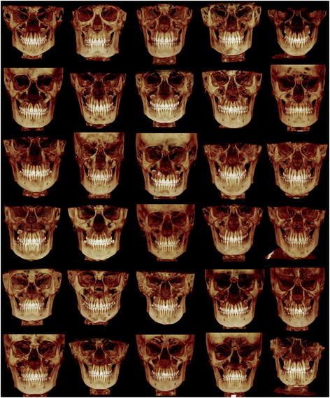
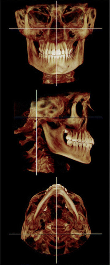
Methods: Thirty consecutive Class I patients (mean age 14.32 years, SD 1.67) meeting the inclusion criteria were analyzed by three-dimensional cone-beam computed tomography (CBCT). Dental, maxillary, mandibular, and cranial base variables were measured with Dolphin 3D. CBCT analysis consisted of the localization of 34 anatomical landmarks. All reference points were digitized in 3D and analyzed using 67 skeletal and dental measurements. Student's t tests for paired samples were used with a significance level of p < 0.05.
Results: Minor right-left discrepancies were noted in all planes. The most anterior point of the glenoid fossa and most condylar points were positioned more superior and lateral on the right side, compared to the left side. Porion was also located more superiorly on the right side relative to the left side. The posterior nasal spine was found to be located to the right of the midsagittal plane. Slight dental midline discrepancies were found, and the dental arch lengths were slightly longer on the left side compared to the right. The height of the ramus, in both 3D and 2D, and the inclination of the ramus were greater on the right than that on the left side.
Conclusions: The findings of this study suggest minor asymmetries exist and are likely a common occurrence in the normal human craniofacial complex. Additionally, a natural compensatory mechanism may exist which controls the size and shape of specific tissues in order to maintain functional symmetry.
Figures
References
-
- Proffit WR, White RP, Sarver DM. Contemporary treatment of dentofacial deformity. Mosby, Maryland Heights, Missouri; 2003.
Publication types
MeSH terms
LinkOut - more resources
Full Text Sources
Other Literature Sources
