. 2014 Jun 30;211(7):1449-64.
doi: 10.1084/jem.20132126. Epub 2014 Jun 16.

Cardiac fibroblasts mediate IL-17A-driven inflammatory dilated cardiomyopathy

Affiliations

Cardiac fibroblasts mediate IL-17A-driven inflammatory dilated cardiomyopathy

Lei Wu et al. J Exp Med. .

Abstract

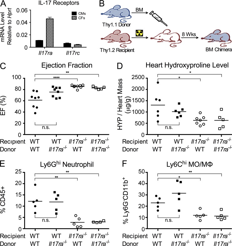
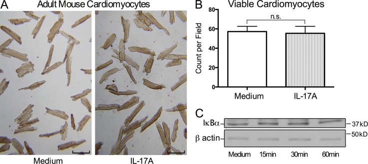
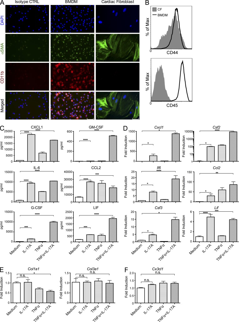
Inflammatory dilated cardiomyopathy (DCMi) is a major cause of heart failure in individuals below the age of 40. We recently reported that IL-17A is required for the development of DCMi. We show a novel pathway connecting IL-17A, cardiac fibroblasts (CFs), GM-CSF, and heart-infiltrating myeloid cells with the pathogenesis of DCMi. Il17ra(-/-) mice were protected from DCMi, and this was associated with significantly diminished neutrophil and Ly6Chi monocyte/macrophage (MO/MΦ) cardiac infiltrates. Depletion of Ly6Chi MO/MΦ also protected mice from DCMi. Mechanistically, IL-17A stimulated CFs to produce key chemokines and cytokines that are critical downstream effectors in the recruitment and differentiation of myeloid cells. Moreover, IL-17A directs Ly6Chi MO/MΦ in trans toward a more proinflammatory phenotype via CF-derived GM-CSF. Collectively, this IL-17A-fibroblast-GM-CSF-MO/MΦ axis could provide a novel target for the treatment of DCMi and related inflammatory cardiac diseases.


Figures

Figure 1.
Figure 1.
Il17ra−/− mice are protected from DCMi. EAM and DCMi were induced in WT and Il17ra−/− mice. On days 0 and 7, mice received s.c. immunizations of 100 µg MyHCα614-629 peptide emulsified in CFA supplemented to 5 mg/ml of heat-killed M. tb strain H37Ra. On day 0, mice also received 500 ng pertussis toxin i.p. (A) Mice were sacrificed 21 d after immunization. EAM was scored using H&E staining as described in Materials and methods. Data are representative of 3 independent experiments. Data points represent individual mice. Horizontal bars represent mean. Data are analyzed by Mann-Whitney U test. n.s. = not significant. (B–G) 63 d after immunization, naive and immunized mice underwent echocardiography and were sacrificed. (B) Representative M-Mode echocardiography of naive, WT, and Il17ra−/− mice. (C) Ejection fraction (%) of naive, WT, and Il17ra−/− mice by echocardiography. Dotted line marks 60%, the threshold for severe DCMi. (D) Heart weight/body weight ratio (‰). (E) Cardiac hydroxyproline assay normalized to heart weight. (F) Representative histopathology of Il17ra−/− and WT mice hearts showing Masson’s trichrome blue staining. Fibrotic tissue was stained blue. Bars, 1 mm. (G) Cardiac fibrosis in Il17ra−/− and WT mice scored using Masson’s trichrome blue staining. (B–G) Data are representative of 3 independent experiments. Data points represent individual mice. Horizontal bars represent mean. Data were analyzed by one-way ANOVA followed by Tukey’s post-test. *, P < 0.05; **, P < 0.01; ****, P < 0.0001.
Figure 2.
Figure 2.
IL-17RA deficiency alters the composition of heart-infiltrating cells. EAM and DCMi were induced in WT and Il17ra−/− mice. Mice were sacrificed 21 d after immunization. (A–F) The composition of heart-infiltrating inflammatory cells was analyzed by flow cytometry. (A) Representative gating of heart-infiltrating myeloid cells. (B) Total cell number of intracardiac CD45+ leukocytes in WT and Il17ra−/− mice. (C) Intracardiac Ly6Ghi neutrophils as a proportion of total CD45+ leukocytes. (D) Intracardiac Ly6GCD11b+ MO/MΦs as a proportion of total CD45+ leukocytes. (E) Ly6Chi MO/MΦs as a proportion of Ly6GCD11b+ population. (F) Ly6Chi to Ly6Clo MO/MΦ ratio. (G–I) The composition of splenocytes was analyzed by flow cytometry with gating similar to A. (G) Ly6Chi monocytes as a proportion of total Ly6GCD11cCD11b+F4/80 population in the spleen. (H) Ly6Chi to Ly6Clo monocyte ratio in the spleen. (I) Ly6Ghi neutrophils as a proportion of total CD45+ leukocytes in the spleen. For bar graphs, data are representative of 3 independent experiments. Data points represent individual mice. Horizontal bars represent mean. Data are analyzed by unpaired two-tailed Student’s t test. **, P < 0.01; ****, P < 0.0001; n.s. = not significant.
Figure 3.
Figure 3.
Transcriptomes and functions of intracardiac Ly6Chi and Ly6Clo MO/MΦs. (A) EAM and DCMi were induced in WT mice. Mice were sacrificed 21 d after immunization. Ly6GCD11b+Ly6Chi (Ly6Chi) and Ly6GCD11b+Ly6Clo (Ly6Clo) MO/MΦ populations were isolated from mouse hearts by FACS. mRNA levels were determined by qPCR, normalized to housekeeping gene Hprt. Detailed data are shown in Table 1. For each individual gene, the ratio of its expression in Ly6Chi population versus that in Ly6Clo was calculated and plotted in log scale. Ratio greater than one indicates the gene was up-regulated in Ly6Chi MO/MΦs (orange), and ratio smaller than one indicates that the gene was up-regulated in Ly6Clo MO/MΦs (blue). Data are representative of 2 independent experiments. (B–E) EAM and DCMi were induced in WT mice. On days 14, 16, 18, and 20, mice were injected i.v. with 250 µl PBS (control), PBS-loaded liposome (PBS liposome), or clodronate-loaded liposome (clodronate). Mice were sacrificed 21 d after immunization. The composition of heart-infiltrating cells was analyzed by flow cytometry. Data are representative of 2 independent experiments. (B) Ly6Chi MO/MΦs as a proportion of Ly6GCD11b+ population. (C) Ly6Chi to Ly6Clo MO/MΦ ratio. (D) Intracardiac Ly6GCD11b+ MO/MΦs as a proportion of total CD45+ leukocytes. (E) Intracardiac Ly6Ghi neutrophils as a proportion of total CD45+ leukocytes. (F–H) EAM and DCMi were induced in WT mice. From days 14 to 35, mice were injected i.v. every other day with 250 µl PBS (control), PBS-loaded liposome (PBS liposome), or clodronate-loaded liposome (clodronate). 63 d after immunization, mice underwent echocardiography and were sacrificed. Data are representative of 2 independent experiments. (F) Ejection fraction (%) by echocardiography. Dash line marks 60%, the threshold for severe DCMi. (G) Heart weight/body weight ratio (‰). (H) Cardiac hydroxyproline assay normalized to heart weight. For bar graphs, data points represent individual mice. Horizontal bars represent mean. Data were analyzed by one-way ANOVA followed by Tukey’s post-test. *, P < 0.05; **, P < 0.01; ***, P < 0.001; n.s. = not significant.
Figure 4.
Figure 4.
IL-17RA signaling to cardiac-resident cells is required for the development of DCMi. (A) Primary adult mouse CMs and CFs were isolated from naive WT mice. mRNA of Il17ra and Il17rc were detected by real-time qPCR. Data are representative of 2 independent experiments. Data are shown as mean + SEM of three replicates. (B) Schematic of the generation of BM chimeras. BMs were transferred from WT or Il17ra−/− Thy1.1 donor into lethally irradiated Il17ra−/− or WT Thy1.2 recipients. EAM and DCMi were induced 8 wk after transfer. (C and D) 63 d after immunization, chimeric mice underwent echocardiography and were sacrificed. Data are representative of 4 independent experiments. (C) Ejection fraction (%) of BM chimeras with depicted genotypes. (D) Cardiac hydroxyproline assay normalized to heart weight. (E and F) 21 d after immunization, chimeric mice were sacrificed, and their heart-infiltrating cells were analyzed by flow cytometry. Data are representative of 3 independent experiments. (E) Intracardiac Ly6Ghi neutrophil as a proportion of CD45+ leukocytes in BM chimeras with depicted genotype. (F) Ly6Chi MO/MΦs as a proportion of Ly6GCD11b+ population. For bar graphs, data points represent individual mice. Horizontal bars represent mean. Data are analyzed by two-way ANOVA (genotype of donor vs. genotype of recipient) followed by Tukey’s post-test. *, P < 0.05; **, P < 0.01; ****, P < 0.0001; n.s. = not significant.
Figure 5.
Figure 5.
IL-17A has no significant effects on adult mouse CMs in vitro. (A and B) Primary adult mouse CMs were cultured with or without 100 ng/ml rIL-17A for 24 h. Data are representative of 3 independent experiments. (A) Bright field microscopy showed cell morphology and viability of rIL-17A–treated and control CMs. Bars, 100 µm. (B) Viable CMs were counted in 5 different fields. Data are shown as mean + SEM and analyzed by unpaired two-tailed Student’s t test. n.s. = not significant. (C) Primary adult CMs were stimulated with 100 ng/ml rIL-17A for 15, 30, or 60 min. Cells were lysed and probed by Western blotting for IκBα and β actin as control. Data are representative of 2 independent experiments.
Figure 6.
Figure 6.
IL-17A stimulates the production of myeloid chemokines and cytokines in adult CFs in vitro. (A) Primary adult mouse CFs from naive WT mice and BMDMs were cultured on chamber slides. Immunofluorescence microscopy shows staining for α-SMA (green) and CD11b (red). BMDMs were stained with isotype-matched antibodies as isotype control. Data are representative of 2 independent experiments. Bars, 100 µM. (B) The expression of surface markers CD44 and CD45 in CF (filled) and BMDM (open) cultures were analyzed by flow cytometry. Levels of expression on all 7-AAD–negative viable cells from respective cultures were plotted on histograms. Data are representative of 2 independent experiments. (C) CFs from naive WT mice were cultured with 100 ng/ml rIL-17A, 5 ng/ml rTNF, or two cytokines combined for 24 h. Supernatants were collected after culture, and the levels of CXCL1, CCL2, GM-CSF, G-CSF, IL-6, and LIF were measured by ELISA. (D–F) CFs from naive WT mice were cultured with 100 ng/ml rIL-17A, 5 ng/ml rTNF, or two cytokines combined for 6 h. RNA were isolated from CFs and the mRNA levels of Cxcl1, Ccl2, Csf2, Csf3, Il6, and Lif (D), Col1a1 and Col3a1 (E), and Cx3cl1 (F) were determined by real-time qPCR and normalized to housekeeping gene Hprt. (C–F) Data are representative of 3 independent experiments. Data are shown as mean + SEM of 3 replicates and analyzed by one-way ANOVA followed by Tukey’s post-test. *, P < 0.05; **, P < 0.01; ***, P < 0.001; ****, P < 0.0001; n.s. = not significant.
Figure 7.
Figure 7.
CFs react to IL-17A to produce proinflammatory cytokines and chemokines in vivo. EAM and DCMi were induced in WT and Il17ra−/− mice. Mice were sacrificed 21 d after immunization. CD45CD34+CD146+CD44hiCD31 CFs and CD45CD34+CD146+CD44loCD31hi ECs were isolated from mouse hearts by FACS. (A) Representative gating of CFs and ECs from viable singlets. (B) mRNA levels of Cxcl1, Csf2, Csf3, and Il6 in CFs from WT and Il17ra−/− mice were determined by qPCR and normalized to housekeeping gene Hprt. (C) mRNA levels of Ccl2, Cxcl1, Csf2, Csf3, Il6, and Lif in CFs and ECs from WT mice were determined by qPCR and normalized to housekeeping gene Hprt. For bar graphs, data are representative of 2 independent experiments. Data are shown as mean + SEM of 3 replicates and analyzed by unpaired two-tailed Student’s t test. *, P < 0.05; ***, P < 0.001; ****, P < 0.0001; n.s. = not significant.
Figure 8.
Figure 8.
IL-17A drives the differentiation of monocytes in trans through CFs and GM-CSF, but has no effect in the balance of Ly6Chi and Ly6Clo populations. (A) Ly6GCD11cCD11b+F4/80Ly6Chi spleen monocytes were isolated from naive Il17ra−/− mice by FACS, and co-cultured with primary adult CFs from naive WT mice for 48 h under various conditions as depicted. mRNAs were isolated from FACS-sorted monocytes in the end of co-culture. The mRNA levels of Il1b, Il6, Il12a, Nos2, and Il10 were measured by real-time qPCR, and normalized to Hprt. Data are representative of 3 independent experiments. (B) Ly6GCD11cCD11b+F4/80Ly6Chi spleen monocytes were isolated from naive Il17ra−/− mice by FACS, and co-cultured with primary adult CFs from naive Il17ra−/− mice for 48 h with or without 100 ng/ml rIL-17A. mRNAs were isolated from FACS-sorted monocytes in the end of co-culture. The mRNA levels of Il1b, Il6, Il12a, Nos2, and Il10 were measured by real-time qPCR and normalized to Hprt. (C) Ly6GCD11cCD11b+F4/80 splenic monocytes with mixed Ly6Chi and Ly6Clo populations were isolated from naive WT mice by FACS, and stimulated with 100 ng/ml rIL-17A for 48 h. Ly6Chi monocyte as a proportion of total was analyzed by flow cytometry. (D) Ly6GCD11cCD11b+F4/80 splenic monocytes with mixed Ly6Chi and Ly6Clo populations were isolated from naive Il17ra−/− mice by FACS, and cultured alone (medium), with WT CFs (CF), or with WT CF and rIL-17A (CF+IL-17A) for 48 h. Ly6Chi monocyte as a proportion of total was analyzed by flow cytometry. (B–D) Data are representative of 2 independent experiments. (A–D) Data are shown as mean + SEM of 3 replicates and analyzed by and one-way ANOVA followed by Tukey’s post-test (A and D), or unpaired two-tailed Student’s t test (B and C). *, P < 0.05; **, P < 0.01; ***, P < 0.001; ****, P < 0.0001; n.s. = not significant.
Figure 9.
Figure 9.
IL-17A and GM-CSF drive cardiac infiltration of Ly6Chi MO/MΦs into proinflammatory phenotype in vivo. (A and B) EAM and DCMi were induced in WT and Il17ra−/− mice. Mice were sacrificed 21 d after immunization. (A) Ly6GCD11b+Ly6Chi MO/MΦs were isolated from mouse hearts by FACS. (B) Ly6GCD11cCD11b+F4/80Ly6Chi monocytes were isolated from mouse spleens by FACS. (C) EAM and DCMi were induced in WT mice. Mice were injected i.p. with PBS or 0.5 µg rGM-CSF, 36 and 12 h before sacrifice at day 21. Ly6GCD11b+Ly6Chi MO/MΦs were isolated from mouse hearts by FACS. (A–C) mRNA levels of Il1b and Il6 were determined by qPCR and normalized to housekeeping gene Hprt. Data are representative of 2 independent experiments. Data are shown as mean + SEM of 3 replicates and analyzed by unpaired two-tailed Student’s t test. *, P < 0.05; n.s. = not significant.

References

    1. Aretz H.T., Billingham M.E., Edwards W.D., Factor S.M., Fallon J.T., Fenoglio J.J., Jr, Olsen E.G., Schoen F.J. 1987. Myocarditis. A histopathologic definition and classification. Am. J. Cardiovasc. Pathol. 1:3–14 - PubMed
    1. Baldeviano G.C., Barin J.G., Talor M.V., Srinivasan S., Bedja D., Zheng D., Gabrielson K., Iwakura Y., Rose N.R., Cihakova D. 2010. Interleukin-17A is dispensable for myocarditis but essential for the progression to dilated cardiomyopathy. Circ. Res. 106:1646–1655 10.1161/CIRCRESAHA.109.213157 - DOI - PubMed
    1. Barin J.G., Rose N.R., Ciháková D. 2012. Macrophage diversity in cardiac inflammation: a review. Immunobiology. 217:468–475 10.1016/j.imbio.2011.06.009 - DOI - PMC - PubMed
    1. Bessich J.L., Nymon A.B., Moulton L.A., Dorman D., Ashare A. 2013. Low levels of insulin-like growth factor-1 contribute to alveolar macrophage dysfunction in cystic fibrosis. J. Immunol. 191:378–385 10.4049/jimmunol.1300221 - DOI - PMC - PubMed
    1. Blyszczuk P., Behnke S., Lüscher T.F., Eriksson U., Kania G. 2013. GM-CSF promotes inflammatory dendritic cell formation but does not contribute to disease progression in experimental autoimmune myocarditis. Biochim. Biophys. Acta. 1833:934–944 10.1016/j.bbamcr.2012.10.008 - DOI - PubMed

Publication types

MeSH terms

Substances