. 2014 Jul 29;34(4):e00126.
doi: 10.1042/BSR20140031.

Using real-time impedance-based assays to monitor the effects of fibroblast-derived media on the adhesion, proliferation, migration and invasion of colon cancer cells

Affiliations

Using real-time impedance-based assays to monitor the effects of fibroblast-derived media on the adhesion, proliferation, migration and invasion of colon cancer cells

Catríona M Dowling et al. Biosci Rep. .

Abstract

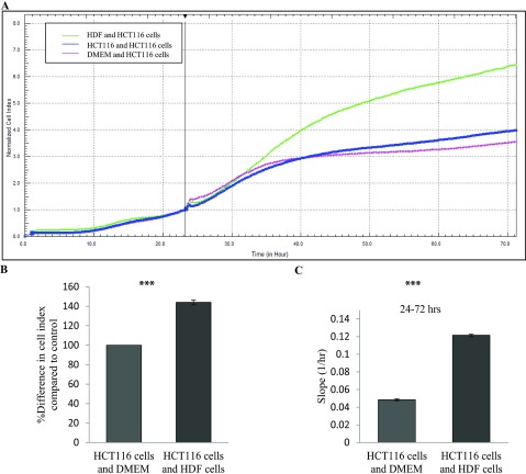
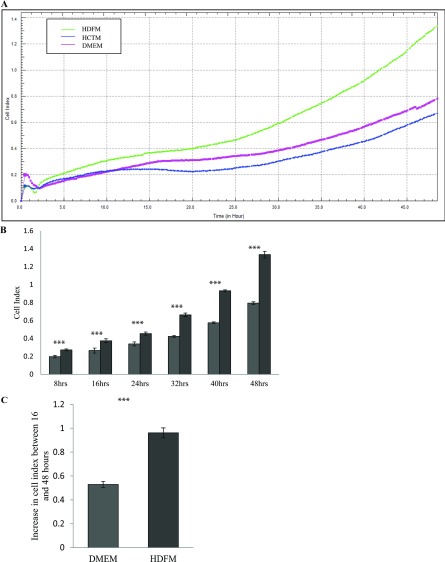
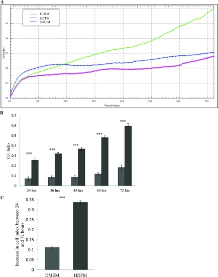
Increasing our knowledge of the mechanisms regulating cell proliferation, migration and invasion are central to understanding tumour progression and metastasis. The local tumour microenvironment contributes to the transformed phenotype in cancer by providing specific environmental cues that alter the cells behaviour and promotes metastasis. Fibroblasts have a strong association with cancer and in recent times there has been some emphasis in designing novel therapeutic strategies that alter fibroblast behaviour in the tumour microenvironment. Fibroblasts produce growth factors, chemokines and many of the proteins laid down in the ECM (extracellular matrix) that promote angiogenesis, inflammation and tumour progression. In this study, we use a label-free RTCA (real-time cell analysis) platform (xCELLigence) to investigate how media derived from human fibroblasts alters cancer cell behaviour. We used a series of complimentary and novel experimental approaches to show HCT116 cells adhere, proliferate and migrate significantly faster in the presence of media from human fibroblasts. As well as this, we used the xCELLigence CIM-plates system to show that HCT116 cells invade matrigel layers aggressively when migrating towards media derived from human fibroblasts. These data strongly suggest that fibroblasts have the ability to increase the migratory and invasive properties of HCT116 cells. This is the first study that provides real-time data on fibroblast-mediated migration and invasion kinetics of colon cancer cells.


Figures

Figure 1
Figure 1. Optimizing cell number
HCT116 cells were seeded at numbers ranging from 20 000, 30 000 and 40 000 in each well of an E-plate and the cells were automatically monitored every 30 seconds over 24 h. Results were expressed as a CI value. (A) Representative graph from xCELLigence system comparing the growth curve of HCT116 cells at 20 000 cells (purple line), 30 000 cells (green line) and 40 000 cells (orange line) (n=3). (B) Shown here is the rate of proliferation at the various cell concentrations as determined by analysing the slope of the line between the 6 and 12 h interval.
Figure 2
Figure 2. Effect of HDFM on the rate of proliferation of HCT116 cells
HDFs were incubated with DMEM for 24 h. The media (HDFM) was removed and placed on HCT116 cells. The rate of proliferation was monitored in real-time using the xCELLligence system (n=3). (A) Representative graph comparing the rate of proliferation of HCT116 when incubated with HDFM (green line), HCTM (blue line) or DMEM (pink line). (B) Comparison of the percentage difference in the mean CI between the cells incubated with HDFM or DMEM. (P<0.05, n=3). (C) The rate of proliferation as determined by analysing the slope of the line between the 6 and 12 hour interval (P<0.05, n=3). (D) Representative graph comparing the rate of proliferation of HCT116 when incubated with the two controls HCTM (blue line) or DMEM (pink line). (E) The rate of proliferation of the controls as compared between the 6 and 12 h interval.
Figure 3
Figure 3. Effect of HDFM on the rate of adherence of HCT116 cells
HDFs were incubated with DMEM for 24 h. The media (HDFM) was removed and placed on HCT116 cells. The rate of adherence was monitored between 0 and 3 h, in real-time using the xCELLligence system (n=3). (A) Representative graph comparing the rate of adherence of HCT116 when incubated with HDFM (green line), HCTM (blue line) or DMEM (pink line). (B) Comparison of the percentage difference in the mean CI between the cells incubated with HDFM or DMEM (P<0.05, n=3). (C) Representative graph comparing the rate of adherence of HCT116 when incubated with the two controls HCTM (blue line) or DMEM (pink line). (D) The CI of the two controls measured at 3 h.
Figure 4
Figure 4. Effect of co-culturing HDF cells with HCT116 cells
HCT116 cells were seeded in the E-plate 16. 24 h later an E-plate insert containing HDF cells was placed into the E-plate 16. The rate of proliferation was monitored in real-time using the xCELLigence system. (A) Representative graph comparing the rate of proliferation of HCT116 co-cultured with HDF cells (green line), HCT116 cells (blue line) or DMEM (pink line). (B) Comparison of the percentage difference in the mean CI between the cells co cultured with HDF cells or DMEM (P<0.05, n=1). (C) The rate of proliferation as determined by analysing the slope of the line between the 24 and 72 h interval (P<0.05).
Figure 5
Figure 5. Effect of HDFM on the rate of migration of HCT116 cells
HDF cells were incubated with DMEM for 24 h. The media (HDFM) was removed and placed in the LC of the CIM -plate with HCT116 cells in the UC (see the Method section). The rate of migration was monitored in real-time using the xCELLigence system (n=3). (A) Representative graph comparing the rate of migration of HCT116 towards HDFM (green line), HCTM (blue line) or DMEM (pink line). (B) Comparison of the CI between the cells migrating towards HDFM or DMEM, at 8, 16, 24, 32, 40 and 48 h (P<0.05, n=3). (C) The change in CI between 16 and 48 h (P<0.05, n=3).
Figure 6
Figure 6. Effect of HDFM on the rate of invasion of HCT116 cells
HDF cells were incubated DMEM for 24 h. The media (HDFM) was removed and placed in the LC of the CIM-plate with HCT116 cells in the UC. The UC of the CIM-plate was coated in matrigel. The rate of invasion was monitored in real-time using the xCELLigence system (n=3). (A) Representative graph comparing the rate of invasion of HCT116 cells as they move towards HDFM (green line), HCTM (blue line) or DMEM (pink line). (B) Comparison of CI between the cells invading the matrigel layer towards HDFM or DMEM, at 24, 36, 48, 60 and 72 h (P<0.05, n=3). (C) The change in CI between 24 and 72 h (P<0.05, n=3).

References

    1. Hanahan D., Weinberg R. A. Hallmarks of cancer: the next generation. Cell. 2011;144:646–674. doi: 10.1016/j.cell.2011.02.013. - DOI - PubMed
    1. Liotta L. A., Kohn E. C. The microenvironment of the tumour-host interface. Nature. 2001;411:375–379. doi: 10.1038/35077241. - DOI - PubMed
    1. Mueller M. M., Fusenig N. E. Friends or foes-bipolar effects of the tumour stroma in cancer. Nat. Rev. Cancer. 2004;4:839–849. doi: 10.1038/nrc1477. - DOI - PubMed
    1. De Wever O., Mareel M. Role of tissue stroma in cancer cell invasion. J. Pathol. 2003;200:429–447. doi: 10.1002/path.1398. - DOI - PubMed
    1. Feng S., Dakhova O., Creighton C. J., Ittmann M. Endocrine fibroblast growth factor FGF19 promotes prostate cancer progression. Cancer Res. 2013;73:2551–2562. doi: 10.1158/0008-5472.CAN-12-4108. - DOI - PMC - PubMed

Publication types