. 2015 May;41(5):1353-64.
doi: 10.1002/jmri.24678. Epub 2014 Jun 17.

Motion artifact reduction in pediatric diffusion tensor imaging using fast prospective correction

Affiliations

Motion artifact reduction in pediatric diffusion tensor imaging using fast prospective correction

A Alhamud et al. J Magn Reson Imaging. 2015 May.

Abstract

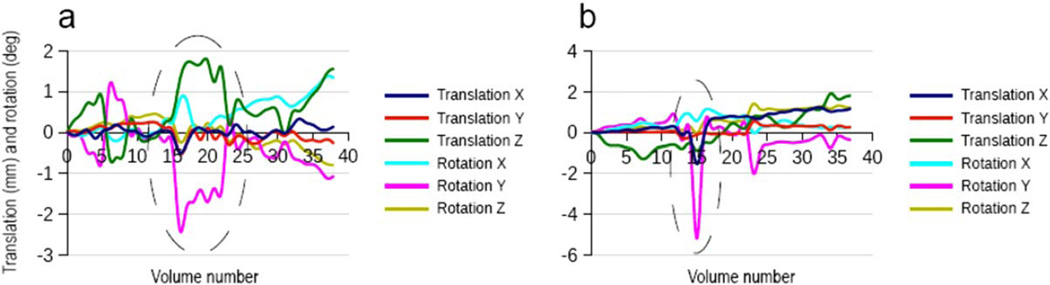
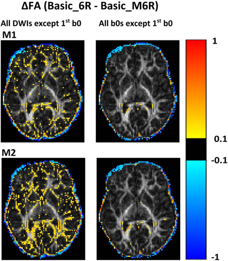
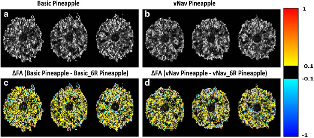
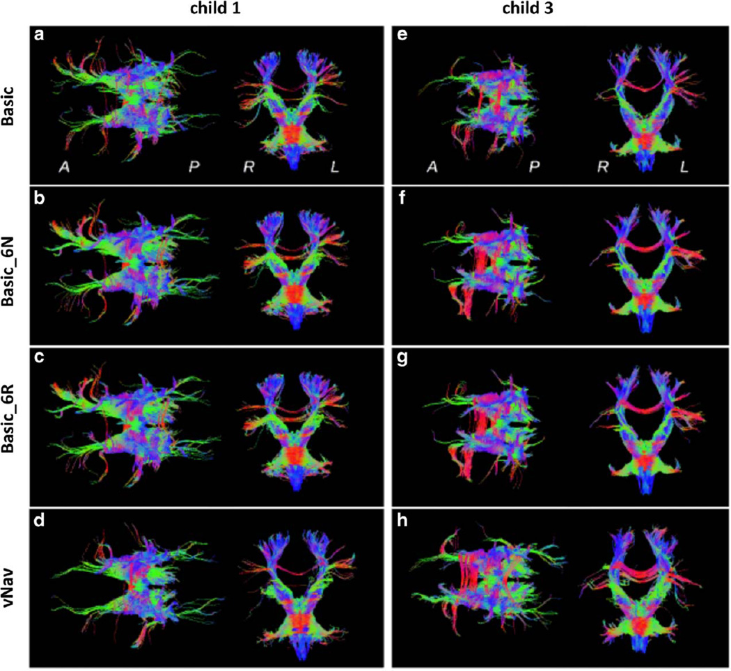
Purpose: To evaluate the patterns of head motion in scans of young children and to examine the influence of corrective techniques, both qualitatively and quantitatively. We investigate changes that both retrospective (with and without diffusion table reorientation) and prospective (implemented with a short navigator sequence) motion correction induce in the resulting diffusion tensor measures.

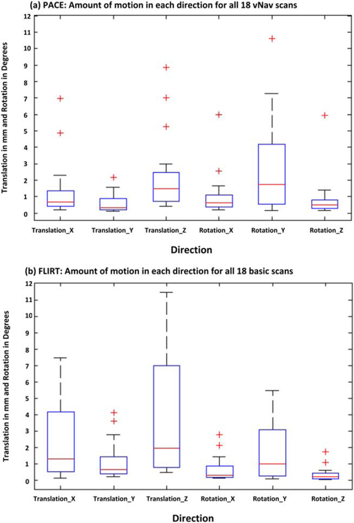
Materials and methods: Eighteen pediatric subjects (aged 5-6 years) were scanned using 1) a twice-refocused, 2D diffusion pulse sequence, 2) a prospectively motion-corrected, navigated diffusion sequence with reacquisition of a maximum of five corrupted diffusion volumes, and 3) a T1 -weighted structural image. Mean fractional anisotropy (FA) values in white and gray matter regions, as well as tractography in the brainstem and projection fibers, were evaluated to assess differences arising from retrospective (via FLIRT in FSL) and prospective motion correction. In addition to human scans, a stationary phantom was also used for further evaluation.

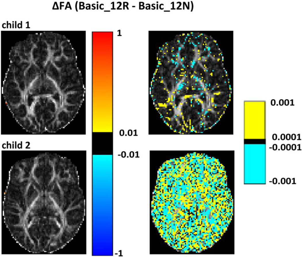
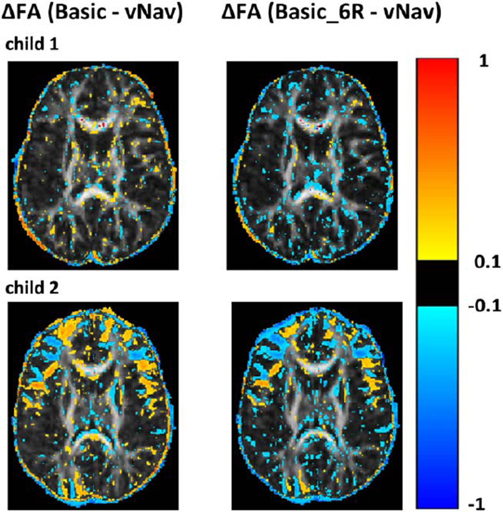
Results: In several white and gray matter regions retrospective correction led to significantly (P < 0.05) reduced FA means and altered distributions compared to the navigated sequence. Spurious tractographic changes in the retrospectively corrected data were also observed in subject data, as well as in phantom and simulated data.

Conclusion: Due to the heterogeneity of brain structures and the comparatively low resolution (∼2 mm) of diffusion data using 2D single shot sequencing, retrospective motion correction is susceptible to distortion from partial voluming. These changes often negatively bias diffusion tensor imaging parameters. Prospective motion correction was shown to produce smaller changes.

Keywords: diffusion tensor imaging (DTI); navigated diffusion sequence (vNav); prospective motion correction; retrospective motion correction; tractography.


Figures

Figure 1
Figure 1
Representative displacements in x-, y-, and z-directions, and rotations around each axis for two children who moved during scanning, as estimated by PACE during the navigated sequence (vNav). a: An example of continuous movement over several volumes. b: Abrupt or fast movement "spikes." In the scanner coordinate system, these axes correspond to: x = anterior–posterior (AP), y = left–right (LR), and z = superior–inferior (SI) directions.
Figure 2
Figure 2
Boxplots showing the amount of motion in each direction (translation and rotation in image space; outliers shown denoted by red "+") for all 18 children as estimated using PACE during the vNav acquisitions in (a) and using FSL-FLIRT for the basic acquisitions in (b). The amount of motion was computed as the difference between the maximum and minimum displacement and rotation for each child. In the scanner coordinate system, axes correspond to: x = AP, y = LR, and z = SI. Children in this study predominantly moved along the z-direction and rotated their heads around the y-axis, which corresponds to nodding motion. [Color figure can be viewed in the online issue, which is available at wileyonlinelibrary.com.]
Figure 3
Figure 3
Voxelwise differences for two children (child 1 and child 2 in Table 1) between FA maps generated for retrospectively motion-corrected data with (Basic_12R) and without (Basic_12N) rotation of the diffusion table superimposed on the basic_12R FA maps. FA differences are shown using two different scales: the left column shows differences on the scale 0.01 < |ΔFA| < 1, and the right on the scale 0.0001 < |ΔFA| < 0.001. Child 2 moved more than child 1.
Figure 4
Figure 4
Voxelwise differences for two children (child 1 and child 2 in Table 1) between FA maps for the basic and vNav acquisitions on the left, and between the basic acquisition after retrospective motion correction with 6DOF and rotation of the gradient table (Basic_6R) and the vNav acquisition on the right. FA differences are overlaid on the vNav FA maps. All FA maps were coregistered to subject T1 space prior to subtraction.
Figure 5
Figure 5
Map of voxelwise differences in FA between the basic acquisition of one child (Basic_6R) and the FA map generated after applying the false transformation matrices M1 (top) and M2 (bottom), respectively, to the same data (Basic_M6R). Preprocessing for all FA maps included retrospective motion correction using 6DOF and rotation of the diffusion table. For the images on the left, the transformations were applied to all the volumes except the first B0 volume, while for the images on the right the transformation was applied only to the 2nd, 3rd, and 4th B0 volume images. Differences are overlaid on the FA map of the retrospectively motion-corrected basic scan.
Figure 6
Figure 6
a,b: FA maps for three slices of a pineapple acquired using the basic and vNav sequences, respectively. c: The voxelwise differences in FA (ΔFA) between FA values in ‘a’ before and after motion correction with 6DOF and rotation of the diffusion table (Basic Pineapple – Basic_6R Pineapple). d: The same for data acquired using the vNav sequence (vNav Pineapple – vNav_6R Pineapple). For both sequences retrospective motion correction significantly reduced the FA.
Figure 7
Figure 7
Tractography reconstructions for fibers passing through the brainstem for two children. Axial projections (anterior is on the left) and coronal projections (anatomic left is on the right) are viewed from superior and anterior locations, respectively, for various reconstructions: (a,e) Basic scan, no retrospective correction; (b,f) Basic_6N; (c,g) Basic_6R; and (d,h) vNav. Coloration is by local spatial orientation: left–right (red); anterior–posterior (green); superior–inferior (blue).

References

    1. Hasan KM, Walimuni IS, Abid H, et al. Multimodal quantitative magnetic resonance imaging of thalamic development and aging across the human lifespan: implications to neurodegeneration in multiple sclerosis. J Neurosci. 2011;31:16826–16832. - PMC - PubMed
    1. Miller JH, McKinstry RC, Philip JV, Mukherjee P, Neil JJ. Diffusion-tensor MR imaging of normal brain maturation: a guide to structural development and myelination. AJR Am J Roentgenol. 2003;180:851–859. - PubMed
    1. Mukherjee P, Miller JH, Shimony JS, et al. Normal brain maturation during childhood: developmental trends characterized with diffusion-tensor MR imaging. Radiology. 2001;221:349–358. - PubMed
    1. Qiu D, Tan LH, Zhou K, Khong PL. Diffusion tensor imaging of normal white matter maturation from late childhood to young adulthood: voxel-wise evaluation of mean diffusivity, fractional anisotropy, radial and axial diffusivities, and correlation with reading development. Neuroimage. 2008;41:223–232. - PubMed
    1. Schmithorst VJ, Wilke M, Dardzinski BJ, Holland SK. Correlation of white matter diffusivity and anisotropy with age during childhood and adolescence: a cross-sectional diffusion-tensor MR imaging study. Radiology. 2002;222:212–218. - PMC - PubMed

Publication types

LinkOut - more resources