Comparative Study
. 2014 Sep;96(3):473-80.
doi: 10.1189/jlb.4A0214-093RR. Epub 2014 Jun 16.

Characterization of lung inflammation and its impact on macrophage function in aging

Affiliations
Comparative Study

Characterization of lung inflammation and its impact on macrophage function in aging

Cynthia H Canan et al. J Leukoc Biol. 2014 Sep.

Abstract

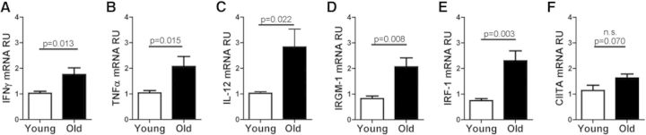
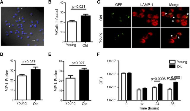
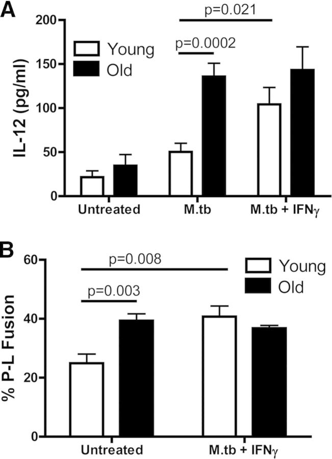
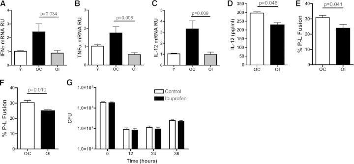
Systemic inflammation that occurs with increasing age (inflammaging) is thought to contribute to the increased susceptibility of the elderly to several disease states. The elderly are at significant risk for developing pulmonary disorders and infectious diseases, but the contribution of inflammation in the pulmonary environment has received little attention. In this study, we demonstrate that the lungs of old mice have elevated levels of proinflammatory cytokines and a resident population of highly activated pulmonary macrophages that are refractory to further activation by IFN-γ. The impact of this inflammatory state on macrophage function was determined in vitro in response to infection with M.tb. Macrophages from the lungs of old mice secreted more proinflammatory cytokines in response to M.tb infection than similar cells from young mice and also demonstrated enhanced M.tb uptake and P-L fusion. Supplementation of mouse chow with the NSAID ibuprofen led to a reversal of lung and macrophage inflammatory signatures. These data indicate that the pulmonary environment becomes inflammatory with increasing age and that this inflammatory environment can be reversed with ibuprofen.

Keywords: M. tuberculosis; age; inflammaging.


Figures

Figure 1.
Figure 1.. Lungs of old mice demonstrate an enhanced inflammatory state, and macrophages isolated from this environment have elevated IFN-γ-induced activation markers.
Relative expression of IFN-γ (A), TNF-α (B), IL-12 (C), IRGM-1 (D), IRF-1 (E), and CIITA (F), as determined by RT-qPCR in old (solid bars) and young (open bars) mice. Whole-lung homogenates (A–C) or purified lung macrophages (D–F) were collected into TRIzol reagent, cDNA was synthesized using the Omniscript RT Kit, and real-time PCR was performed using TaqMan gene expression assays. Data were combined from three independent experiments using five individual young or old mice/experiment (A–C) or from two independent experiments using five individual young or old mice/experiment (D–F). Student's t-test was used to determine statistical significance.
Figure 2.
Figure 2.. Pulmonary macrophages from old mice have increased M.tb uptake and P-L fusion but a defect in intracellular control of bacterial growth.
Pulmonary macrophages isolated from old (solid bars) and young (open bars) mice were adhered to coverslips and incubated with 5:1 GFP-M.tb for 2 h and analyzed by confocal microscopy (A–E). White arrows indicate infected pulmonary macrophages (A). Percentage of cells from both groups found to be infected with GFP-M.tb (B). P-L fusion was observed in macrophages from young and old mice using the lysosomal marker LAMP-1 (C and D) and cathepsin D (E). White arrowhead indicates P-L colocalization events (yellow) between GFP-M.tb (green) and LAMP-1 (red). Macrophages isolated from the lungs of young and old mice were incubated with 5:1 GFP-M.tb for 2 h, and M.tb intracellular growth was assessed at the indicated time-points postinfection (F). CFU was normalized between macrophages from young and old mice at time zero. Colonies were enumerated after 21 days of incubation at 37°C. Data were combined from three independent experiments from pools of five mice. Student's t-test was used to determine statistical significance.
Figure 3.
Figure 3.. IFN-γ fails to boost P-L fusion and cytokine production in response to M.tb infection in pulmonary macrophages from old mice.
Pulmonary macrophages from old (solid bars) and young (open bars) mice were plated onto coverslips and incubated with murine rIFN-γ or left untreated for 16 h. For cytokine production, cells were infected with 5:1 GFP-M.tb for 24 h, and the concentration of IL-12 (A) in the supernatants was analyzed by ELISA. For P-L fusion, cells were infected with 5:1 GFP-M.tb for 2 h, fixed, permeabilized, blocked, and stained for cathepsin D and P-L fusion assessed by confocal microscopy (B). Data were combined from three independent experiments from pools of five mice/experiment. Student's t-test was used to determine statistical significance.
Figure 4.
Figure 4.. Ibuprofen decreases the inflammatory environment and restores macrophage function.
Relative expression of IFN-γ (A), TNF-α (B), and IL-12 (C), as determined by RT-qPCR of whole-lung homogenates from naïve, young mice (Y; open bars) and naïve, old mice on either a control (OC; black bars) or an ibuprofen diet (OI; gray bars). The ΔΔCT method was used to analyze data, normalizing to endogenous 18S rRNA. For cytokine production, cells isolated from old mice on a control (OC; open bars) or an ibuprofen (OI; black bars) diet were infected with 5:1 GFP-M.tb for 24 h, and the secretion of IL-12 was analyzed in supernatants by ELISA (D). For P-L fusion, cells were infected with GFP-M.tb at a MOI of 5:1 for 2 h, fixed, permeabilized, blocked, and stained for LAMP-1 (E) or cathepsin D (F) and P-L fusion events evaluated by confocal microscopy. Macrophages, isolated from the lungs of old mice on a control or an ibuprofen-supplemented diet, were incubated with GFP-M.tb at a MOI of 5:1 for 2 h, and plates were washed with warm DPBS. Bacterial growth (CFUs) at defined time-points postinfection was then assessed by plating lysed macrophages on OADC-supplemented 7H11 agar plates (G). Data were combined from three independent experiments using five individual young, old control, or old ibuprofen mice/experiment (A–C) or three (E and F), two (D), or one (G) independent experiment(s) using pools of five mice/experiment. Student's t-test was used to determine statistical significance.

References

    1. Vasto S., Candore G., Balistreri C. R., Caruso M., Colonna-Romano G., Grimaldi M. P., Listi F., Nuzzo D., Lio D., Caruso C. (2007) Inflammatory networks in ageing, age-related diseases and longevity. Mech. Ageing Dev. 128, 83–91. - PubMed
    1. De Rekeneire N., Peila R., Ding J., Colbert L. H., Visser M., Shorr R. I., Kritchevsky S. B., Kuller L. H., Strotmeyer E. S., Schwartz A. V., Vellas B., Harris T. B. (2006) Diabetes, hyperglycemia, and inflammation in older individuals: the health, aging and body composition study. Diabetes Care 29, 1902–1908. - PubMed
    1. Provinciali M., Cardelli M., Marchegiani F. (2011) Inflammation, chronic obstructive pulmonary disease and aging. Curr. Opin. Pulm. Med. 17 (Suppl. 1), S3–S10. - PubMed
    1. MacNee W. (2011) Aging, inflammation, and emphysema. Am. J. Respir. Crit. Care Med. 184, 1327–1329. - PubMed
    1. Shaw A. C., Joshi S., Greenwood H., Panda A., Lord J. M. (2010) Aging of the innate immune system. Curr. Opin. Immunol. 22, 507–513. - PMC - PubMed

Publication types

MeSH terms