. 2014 May 15;6(3):291-301.
eCollection 2014.

Engineered 3D bioimplants using elastomeric scaffold, self-assembling peptide hydrogel, and adipose tissue-derived progenitor cells for cardiac regeneration

Affiliations

Engineered 3D bioimplants using elastomeric scaffold, self-assembling peptide hydrogel, and adipose tissue-derived progenitor cells for cardiac regeneration

Carolina Soler-Botija et al. Am J Transl Res. .

Abstract

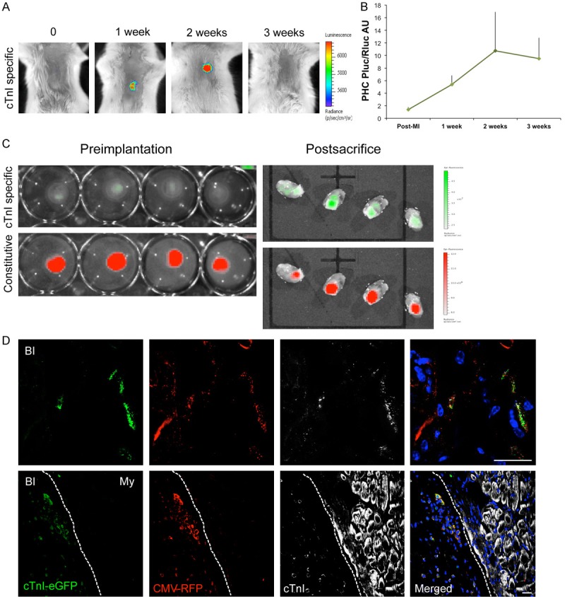
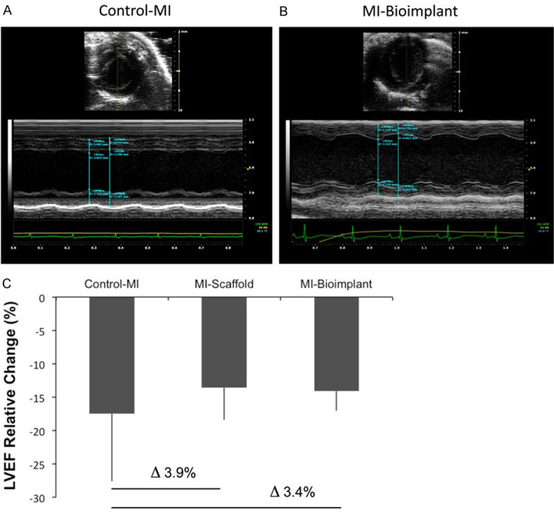
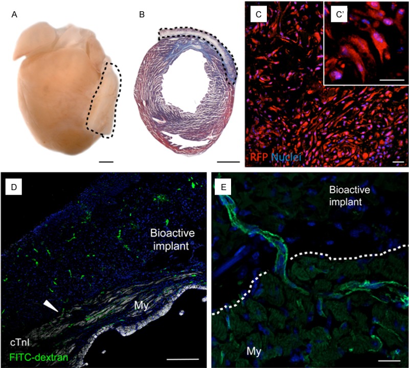
Contractile restoration of myocardial scars remains a challenge with important clinical implications. Here, a combination of porous elastomeric membrane, peptide hydrogel, and subcutaneous adipose tissue-derived progenitor cells (subATDPCs) was designed and evaluated as a bioimplant for cardiac regeneration in a mouse model of myocardial infarction. SubATDPCs were doubly transduced with lentiviral vectors to express bioluminescent-fluorescent reporters driven by constitutively active, cardiac tissue-specific promoters. Cells were seeded into an engineered bioimplant consisting of a scaffold (polycaprolactone methacryloyloxyethyl ester) filled with a peptide hydrogel (PuraMatrix™), and transplanted to cover injured myocardium. Bioluminescence and fluorescence quantifications showed de novo and progressive increases in promoter expression in bioactive implant-treated animals. The bioactive implant was well adapted to the heart, and fully functional vessels traversed the myocardium-bioactive implant interface. Treatment translated into a detectable positive effect on cardiac function, as revealed by echocardiography. Thus, this novel implant is a promising construct for supporting myocardial regeneration.

Keywords: Cardiac regeneration; RECATABI; elastomeric membrane; self-assembling peptide hydrogel; subcutaneous ATDPCs.


Figures

Figure 1
Figure 1
BLI and fluorescent evaluation of cTnI expression and survival of subATDPCs implanted over the mouse infarcted myocardium. A: Representative BLI of labeled subATDPCs within the bioactive implant displays the luciferase signal from the cell differentiation reporter (PLuc-eGFP) that is regulated by the hcTnI promoter. Images of the hcTnI-specific reporter are superimposed on black-and-white dorsal images of the recipient animal. Color bars illustrate relative light intensities from PLuc. B: Histograms of the PLuc/RLuc ratio calculated from photon fluxes recorded via BLI from bioactive implant-treated infarcted animals. C: Representative fluorescence images from subATDPCs within the bioactive implant pre-implantation and post-sacrifice over the excised hearts. Upper images show fluorescence from the cell differentiation reporter (PLuc-eGFP) regulated by hcTnIp (green). Bottom images are representative of constitutive fluorescence from the cell-number reporter (CMVp-RLuc-mRFP1; red). Color bars illustrate the relative fluorescence intensities from eGFP (green) and RFP (red). D: Immunofluorescence staining of mouse heart cross-sections shows the bioactive implant filled with human subATDPCs. Transplanted cells were detected via RFP immunostaining (red), and cTnI expression was detected with anti-eGFP (green) and anti-cTnI (white) antibodies. (BI, bioactive implant; My, myocardium). Scale bars, 20 μm.
Figure 3
Figure 3
Cardiac functional analysis. Representative images and measurements of the LVEF of control-MI (A) and bioactive implant-treated animals (MI-bioimplant) (B) in short axis M-mode. (C) Percentage of LVEF relative change in infarcted groups (control-MI, MI-scaffold, and MI-bioimplant) expressed as the differences between the post-MI and pre-sacrifice values.
Figure 2
Figure 2
Adaptability and vascularization of the bioimplant in the mouse model of MI. A: Representative image of a heart excised from a bioactive implant-treated animal. Dotted lines indicate localization of the implant. Scale bars, 1 mm. B: Masson’s trichrome staining in a heart cross-section reveals the presence of a myocardial scar (blue) and the good adaptability of the bioactive implant. Scale bars, 1 mm. C: Cells inside the bioactive implant three days post-implantation were detected via the constitutively active reporter CMVp-RLuc-mRFP1 (red). C’: Detail of the spindle-shape morphology of subATDPCs. Scale bars, 25 μm. D and E: Functional vessel detection with FITC-dextran (green) in mice treated with subATDPCs. Arrowheads indicate a vessel connection between the mouse myocardium and the bioactive implant in a cell-treated animal. Nuclei were counterstained with Hoechst 33342. Scale bars, 20 μm and 250 μm.

References

    1. Olivetti G, Capasso JM, Meggs LG, Sonnenblick EH, Anversa P. Cellular basis of chronic ventricular remodeling after myocardial infarction in rats. Circ Res. 1991;68:856–869. - PubMed
    1. Anversa P, Leri A, Kajstura J. Cardiac regeneration. J Am Coll Cardiol. 2006;47:1769–1776. - PubMed
    1. Parmacek MS, Epstein JA. Cardiomyocyte renewal. N Engl J Med. 2009;361:86–88. - PMC - PubMed
    1. Bergmann O, Bhardwaj RD, Bernard S, Zdunek S, Barnabé-Heider F, Walsh S, Zupicich J, Alkass K, Buchholz BA, Druid H, Jovinge S, Frisén J. Evidence for cardiomyocyte renewal in humans. Science. 2009;324:98–102. - PMC - PubMed
    1. Genovese J, Cortes-Morichetti M, Chachques E, Frati G, Patel A, Chachques JC. Cell based approaches for myocardial regeneration and artificial myocardium. Curr Stem Cell Res Ther. 2007;2:121–127. - PubMed