

Atypical sulcal anatomy in young children with autism spectrum disorder
- PMID: 24936410
- PMCID: PMC4053636
- DOI: 10.1016/j.nicl.2014.03.008
Atypical sulcal anatomy in young children with autism spectrum disorder
Abstract
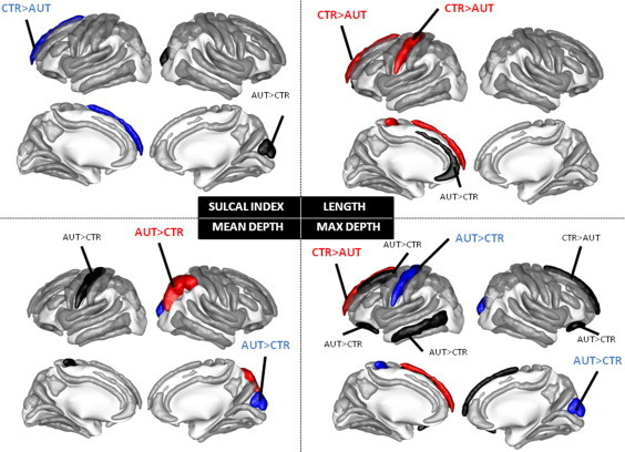
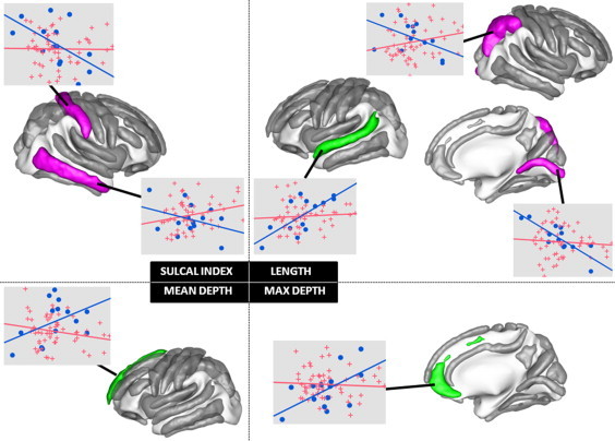
Autism spectrum disorder is associated with an altered early brain development. However, the specific cortical structure abnormalities underlying this disorder remain largely unknown. Nonetheless, atypical cortical folding provides lingering evidence of early disruptions in neurodevelopmental processes and identifying changes in the geometry of cortical sulci is of primary interest for characterizing these structural abnormalities in autism and their evolution over the first stages of brain development. Here, we applied state-of-the-art sulcus-based morphometry methods to a large highly-selective cohort of 73 young male children of age spanning from 18 to 108 months. Moreover, such large cohort was selected through extensive behavioral assessments and stringent inclusion criteria for the group of 59 children with autism. After manual labeling of 59 different sulci in each hemisphere, we computed multiple shape descriptors for each single sulcus element, hereby separating the folding measurement into distinct factors such as the length and depth of the sulcus. We demonstrated that the central, intraparietal and frontal medial sulci showed a significant and consistent pattern of abnormalities across our different geometrical indices. We also found that autistic and control children exhibited strikingly different relationships between age and structural changes in brain morphology. Lastly, the different measures of sulcus shapes were correlated with the CARS and ADOS scores that are specific to the autistic pathology and indices of symptom severity. Inherently, these structural abnormalities are confined to regions that are functionally relevant with respect to cognitive disorders in ASD. In contrast to those previously reported in adults, it is very unlikely that these abnormalities originate from general compensatory mechanisms unrelated to the primary pathology. Rather, they most probably reflect an early disruption on developmental trajectory that could be part of the primary pathology.
Keywords: Autism; MRI; morphometry; sulci.
Figures

References
-
- Alemán-Gómez Y., Janssen J., Schnack H., Balaban E., Pina-Camacho L., Alfaro-Almagro F., Castro-Fornieles J., Otero S., Baeza I., Moreno D., Bargalló N., Parellada M., Arango C., Desco M. The human cerebral cortex flattens during adolescence. Journal of Neuroscience: the Official Journal of the Society for Neuroscience. 2013;33:15004–15010. 24048830 - PMC - PubMed
-
- American Psychiatric Association . Diagnostic and Statistical Manual of Mental Disorders: DSM-IV-TR. Springer; 2000.
Publication types
MeSH terms
LinkOut - more resources
Full Text Sources
Other Literature Sources
Medical
