

Associations between white matter microstructure and amyloid burden in preclinical Alzheimer's disease: A multimodal imaging investigation
- PMID: 24936411
- PMCID: PMC4053642
- DOI: 10.1016/j.nicl.2014.02.001
Associations between white matter microstructure and amyloid burden in preclinical Alzheimer's disease: A multimodal imaging investigation
Abstract
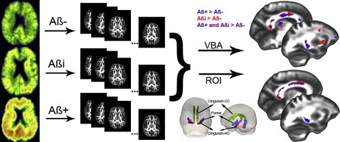
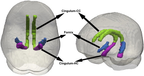
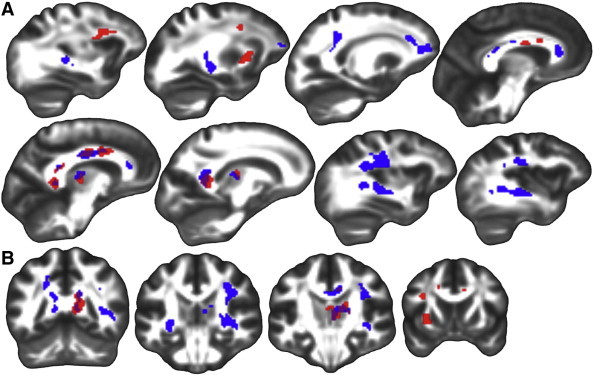
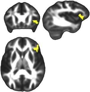
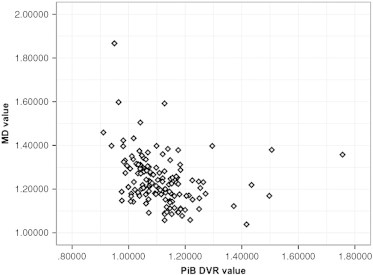
Some cognitively healthy individuals develop brain amyloid accumulation, suggestive of incipient Alzheimer's disease (AD), but the effect of amyloid on other potentially informative imaging modalities, such as Diffusion Tensor Imaging (DTI), in characterizing brain changes in preclinical AD requires further exploration. In this study, a sample (N = 139, mean age 60.6, range 46 to 71) from the Wisconsin Registry for Alzheimer's Prevention (WRAP), a cohort enriched for AD risk factors, was recruited for a multimodal imaging investigation that included DTI and [C-11]Pittsburgh Compound B (PiB) positron emission tomography (PET). Participants were grouped as amyloid positive (Aβ+), amyloid indeterminate (Aβi), or amyloid negative (Aβ-) based on the amount and pattern of amyloid deposition. Regional voxel-wise analyses of four DTI metrics, fractional anisotropy (FA), mean diffusivity (MD), axial diffusivity (Da), and radial diffusivity (Dr), were performed based on amyloid grouping. Three regions of interest (ROIs), the cingulum adjacent to the corpus callosum, hippocampal cingulum, and lateral fornix, were selected based on their involvement in the early stages of AD. Voxel-wise analysis revealed higher FA among Aβ+ compared to Aβ- in all three ROIs and in Aβi compared to Aβ- in the cingulum adjacent to the corpus callosum. Follow-up exploratory whole-brain analyses were consistent with the ROI findings, revealing multiple regions where higher FA was associated with greater amyloid. Lower fronto-lateral gray matter MD was associated with higher amyloid burden. Further investigation showed a negative correlation between MD and PiB signal, suggesting that Aβ accumulation impairs diffusion. Interestingly, these findings in a largely presymptomatic sample are in contradistinction to relationships reported in the literature in symptomatic disease stages of Mild Cognitive Impairment and AD, which usually show higher MD and lower FA. Together with analyses showing that cognitive function in these participants is not associated with any of the four DTI metrics, the present results suggest an early relationship between PiB and DTI, which may be a meaningful indicator of the initiating or compensatory mechanisms of AD prior to cognitive decline.
Keywords: AD risk; ANCOVA, Analysis of Covariance; ANTs, Advanced Normalization Tools; APOE4, apolipoprotein E gene ε4; Alzheimer's disease; Amyloid imaging; Aβ+, amyloid positive; Aβi, amyloid indeterminate; Aβ−, amyloid negative; BET, Brain Extraction Tool; Cingulum–CC, cingulum adjacent to corpus callosum; Cingulum–HC, hippocampal cingulum (projecting to medial temporal lobe); DTI, Diffusion Tensor Imaging; DTI-TK, Diffusion Tensor Imaging Toolkit; DVR, distribution volume ratio; Da, axial diffusivity; Dr, radial diffusivity; FA, fractional anisotropy; FH, (parental) family history; FSL, FMRIB Software Library; FUGUE, FMRIB's utility for geometrically unwarping EPIs; FWE, family wise error; GM, gray matter; HARDI, high angular resolution diffusion imaging; ICBM, International Consortium for Brain Mapping; MD, mean diffusivity; PCC, posterior cingulate cortex; PIB, Pittsburgh compound B; PRELUDE, phase region expanding labeler for unwrapping discrete estimates; RAVLT, Rey Auditory Verbal Learning Test; SPM, Statistical Parametric Mapping; TMT, Trail Making Test; WASI, Wechsler Abbreviated Scale of Intelligence; WM, white matter; WRAP, Wisconsin Registry for Alzheimer's Prevention; WRAT, Wide Range Achievement Test; White matter.
Figures

Similar articles
-
White matter microstructure in late middle-age: Effects of apolipoprotein E4 and parental family history of Alzheimer's disease.Neuroimage Clin. 2014 Apr 21;4:730-42. doi: 10.1016/j.nicl.2014.04.008. eCollection 2014. Neuroimage Clin. 2014. PMID: 24936424 Free PMC article.
-
Cognitively healthy APOE4/4 carriers show white matter impairment associated with serum NfL and amyloid-PET.Neurobiol Dis. 2024 Mar;192:106439. doi: 10.1016/j.nbd.2024.106439. Epub 2024 Feb 15. Neurobiol Dis. 2024. PMID: 38365046
-
Longitudinal changes in microstructural white matter metrics in Alzheimer's disease.Neuroimage Clin. 2016 Dec 16;13:330-338. doi: 10.1016/j.nicl.2016.12.012. eCollection 2017. Neuroimage Clin. 2016. PMID: 28066707 Free PMC article.
-
The role of diffusion tensor imaging and fractional anisotropy in the evaluation of patients with idiopathic normal pressure hydrocephalus: a literature review.Neurosurg Focus. 2016 Sep;41(3):E12. doi: 10.3171/2016.6.FOCUS16192. Neurosurg Focus. 2016. PMID: 27581308 Review.
-
Diffusion tensor imaging in Alzheimer's disease and mild cognitive impairment.Behav Neurol. 2009;21(1):39-49. doi: 10.3233/BEN-2009-0234. Behav Neurol. 2009. PMID: 19847044 Free PMC article. Review.
Cited by
-
Biomarker clusters are differentially associated with longitudinal cognitive decline in late midlife.Brain. 2016 Aug;139(Pt 8):2261-74. doi: 10.1093/brain/aww142. Epub 2016 Jun 20. Brain. 2016. PMID: 27324877 Free PMC article.
-
LOCALIZING DIFFERENTIALLY EVOLVING COVARIANCE STRUCTURES VIA SCAN STATISTICS.Q Appl Math. 2019;77(2):357-398. doi: 10.1090/qam/1522. Epub 2018 Dec 17. Q Appl Math. 2019. PMID: 35125524 Free PMC article.
-
Asymmetric Insular Connectomics Revealed by Diffusion Magnetic Resonance Imaging Analysis of Healthy Brain Development.Brain Connect. 2019 Feb;9(1):2-12. doi: 10.1089/brain.2018.0582. Brain Connect. 2019. PMID: 30501515 Free PMC article.
-
Early brain connectivity alterations and cognitive impairment in a rat model of Alzheimer's disease.Alzheimers Res Ther. 2018 Feb 7;10(1):16. doi: 10.1186/s13195-018-0346-2. Alzheimers Res Ther. 2018. PMID: 29415770 Free PMC article.
-
Cortical microstructural correlates of astrocytosis in autosomal-dominant Alzheimer disease.Neurology. 2020 May 12;94(19):e2026-e2036. doi: 10.1212/WNL.0000000000009405. Epub 2020 Apr 14. Neurology. 2020. PMID: 32291295 Free PMC article.
References
-
- Alexander D.C., Gee J.C. Elastic matching of diffusion tensor images. Comput. Vis. Image Underst. 2000;77:233–250.
-
- Alexander D.C., Pierpaoli C., Basser P.J., Gee J.C. Spatial transformations of diffusion tensor magnetic resonance images. IEEE Trans. Med. Imaging. 2001;20:1131–1139. - PubMed
Publication types
MeSH terms
Substances
Grants and funding
- AG027161/AG/NIA NIH HHS/United States
- T32 CA009206/CA/NCI NIH HHS/United States
- I01 CX000555/CX/CSRD VA/United States
- R01 AG027161/AG/NIA NIH HHS/United States
- T32 AG000213/AG/NIA NIH HHS/United States
- P50 AG033514/AG/NIA NIH HHS/United States
- AG037639/AG/NIA NIH HHS/United States
- R01 AG037639/AG/NIA NIH HHS/United States
- P30 HD003352/HD/NICHD NIH HHS/United States
- I01 CX000165/CX/CSRD VA/United States
- P30 HD003352,/HD/NICHD NIH HHS/United States
- UL1 RR025011/RR/NCRR NIH HHS/United States
- R01 AG021155/AG/NIA NIH HHS/United States
- AG000213/AG/NIA NIH HHS/United States
- AG021155/AG/NIA NIH HHS/United States
LinkOut - more resources
Full Text Sources
Other Literature Sources
Medical
Miscellaneous
