. 2014 Apr 21:4:730-42.
doi: 10.1016/j.nicl.2014.04.008. eCollection 2014.

White matter microstructure in late middle-age: Effects of apolipoprotein E4 and parental family history of Alzheimer's disease

Affiliations

White matter microstructure in late middle-age: Effects of apolipoprotein E4 and parental family history of Alzheimer's disease

Nagesh Adluru et al. Neuroimage Clin. .

Abstract

Introduction: Little is still known about the effects of risk factors for Alzheimer's disease (AD) on white matter microstructure in cognitively healthy adults. The purpose of this cross-sectional study was to assess the effect of two well-known risk factors for AD, parental family history and APOE4 genotype.

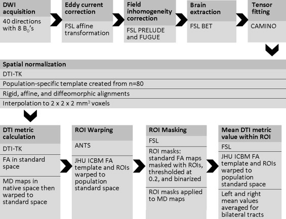
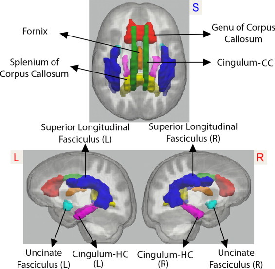
Methods: This study included 343 participants from the Wisconsin Registry for Alzheimer's Prevention, who underwent diffusion tensor imaging (DTI). A region of interest analysis was performed on fractional anisotropy maps, in addition to mean, radial, and axial diffusivity maps, aligned to a common template space using a diffeomorphic, tensor-based registration method. The analysis focused on brain regions known to be affected in AD including the corpus callosum, superior longitudinal fasciculus, fornix, cingulum, and uncinate fasciculus. Analyses assessed the impact of APOE4, parental family history of AD, age, and sex on white matter microstructure in late middle-aged participants (aged 47-76 years).

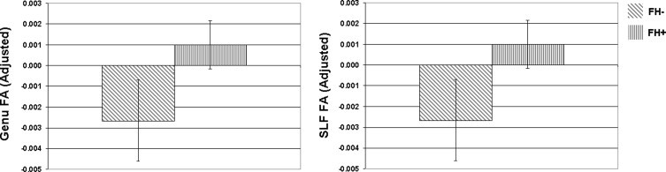
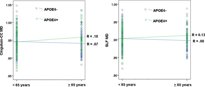
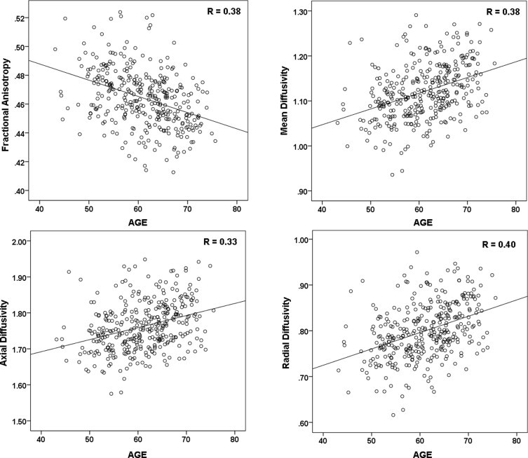
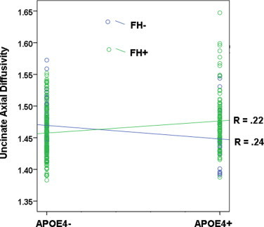
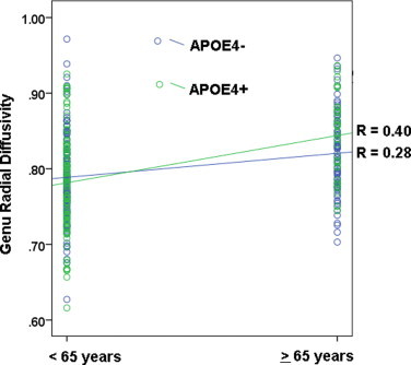
Results: Both APOE4 and parental family history were associated with microstructural white matter differences. Participants with parental family history of AD had higher FA in the genu of the corpus callosum and the superior longitudinal fasciculus. We observed an interaction between family history and APOE4, where participants who were family history positive but APOE4 negative had lower axial diffusivity in the uncinate fasciculus, and participants who were both family history positive and APOE4 positive had higher axial diffusivity in this region. We also observed an interaction between APOE4 and age, whereby older participants (=65 years of age) who were APOE4 carriers, had higher MD in the superior longitudinal fasciculus and in the portion of the cingulum bundle running adjacent to the cingulate cortex, compared to non-carriers. Older participants who were APOE4 carriers also showed higher radial diffusivity in the genu compared to non-carriers. Across all participants, age had an effect on FA, MD, and axial and radial diffusivities. Sex differences were observed in FA and radial diffusivity.

Conclusion: APOE4 genotype, parental family history of AD, age, and sex are all associated with microstructural white matter differences in late middle-aged adults. In participants at risk for AD, alterations in diffusion characteristics-both expected and unexpected-may represent cellular changes occurring at the earliest disease stages, but further work is needed. Higher mean, radial, and axial diffusivities were observed in participants who are more likely to be experiencing later stage preclinical pathology, including participants who were both older and carried APOE4, or who were positive for both APOE4 and parental family history of AD.

Keywords: APOE4; Alzheimer's disease; MRI; age; diffusion tensor imaging; family history; risk factors; sex.


Figures

Fig. 1
Fig. 1
DTI processing stream. Shown here is the diffusion tensor imaging “processing pipeline”. Images underwent eddy current correction, field inhomogeneity correction, skull stripping and tensor fitting using tools from the FMRIB Software Library (FSL). Tensor fitting was performed using the University College London, Camino Diffusion MRI Toolkit. Next, images were normalized utilizing Diffusion Toolkit for DTI analysis (DTI-TK). Advanced Normalization Tools (ANTS) was used to warp the Johns Hopkins International Consortium for Brain Mapping (JHU ICBM) FA template and white matter regions of interest (ROIs) to the study's template space. ROIs were used to extract fractional anisotropy (FA), mean diffusivity (MD), axial diffusivity, and radial diffusivity values from individual subject DTI maps in template space. ROIs are depicted in Fig. 2.
Fig. 2
Fig. 2
Regions of interest. We analyzed fractional anisotropy and mean, axial, and radial diffusivity values extracted from the regions of interest depicted here: genu of the corpus callosum (red), splenium of the corpus callosum (yellow), fornix (orange), superior longitudinal fasciculus, left and right combined (blue), cingulum bundle adjacent to the cingulate gyrus (called cingulum-CC in the manuscript text), left and right combined (green), cingulum projections to the hippocampus (called cingulum-HC in the manuscript text), left and right combined (magenta), and uncinate, left and right combined (blue). L = left, R = right.
Fig. 3
Fig. 3
Effects of parental family history on fractional anisotropy. Participants with parental family history (FH) of AD showed higher mean FA in the genu of the corpus callosum and the superior longitudinal fasciculus (SLF) compared to participants who were family history negative. The bar graph shows the regional mean FA adjusted for age and sex. FA = fractional anisotropy; error bars are standard error of the mean. NB: while means in the genu and SLF appear very similar, they were not exactly the same (the correlation between genu and SLF FA across all participants was strong, R = .43).
Fig. 5
Fig. 5
Interaction between APOE4 and age: mean diffusivity. There was a significant interaction between APOE4 and age, whereby carriers of the APOE4 allele showed higher mean diffusivity (MD) in older age (=65 years) compared to younger age (<65 years). The effect was significant in the cingulum bundle adjacent to the cingulate cortex (cingulum-CC), shown on the left, and the superior longitudinal fasciculus (SLF), shown on the right. Participants who were carriers of the APOE4 allele are represented by green circles.
Fig. 4
Fig. 4
Effect of age. Shown here are the patterns of change in the genu, a region that showed change across all DTI maps. The points represent individual participants. With older age, FA is lower, and mean, axial, and radial diffusivities are higher.
Fig. 6
Fig. 6
Interaction between APOE4 and parental family history of AD. When evaluating the effect of risk for AD on axial diffusivity, we observed a cross-over interaction. Participants who were both APOE4 positive (APOE4+) and family history positive (FH+) showed higher axial diffusivity in the uncinate fasciculus. In contrast participants who were only FH+ showed lower axial diffusivity compared to FH-. The finding suggests that stages of white matter pathology may differ at different levels of risk for AD. Participants with positive family history of AD are represented by green circles.
Fig. 7
Fig. 7
Interaction between APOE4 and age: radial diffusivity. We observed an interaction between APOE4 and age in the genu, where older APOE4 carriers had higher radial diffusivity in the genu compared to APOE4 non-carriers.

References

    1. Abe O., Aoki S., Hayashi N., Yamada H., Kunimatsu A., Mori H., Yoshikawa T., Okubo T., Ohtomo K. Normal aging in the central nervous system: quantitative MR diffusion-tensor analysis. Neurobiology of Aging. 2002;23:433–441. 11959406 - PubMed
    1. Acosta-Cabronero J., Williams G.B., Pengas G., Nestor P.J. Absolute diffusivities define the landscape of white matter degeneration in Alzheimer's disease. Brain: A Journal of Neurology. 2010;133:529–539. 19914928 - PubMed
    1. Adluru N., Zhang H., Fox A.S., Shelton S.E., Ennis C.M., Bartosic A.M., Oler J.A., do Tromp P.M., Zakszewski E., Gee J.C., Kalin N.H., Alexander A.L. A diffusion tensor brain template for rhesus macaques. Neuroimage. 2012;59:306–318. 21803162 - PMC - PubMed
    1. Alexander A.L., Hasan K.M., Lazar M., Tsuruda J.S., Parker D.L. Analysis of partial volume effects in diffusion-tensor MRI. Magnetic Resonance in Medicine: Official Journal of the Society of Magnetic Resonance in Medicine / Society of Magnetic Resonance in Medicine. 2001;45:770–780. 11323803 - PubMed
    1. Alexander A.L., Hurley S.A., Samsonov A.A., Adluru N., Hosseinbor A.P., Mossahebi P., do Tromp P.M., Zakszewski E., Field A.S. Characterization of cerebral white matter properties using quantitative magnetic resonance imaging stains. Brain Connectivity. 2011;1:423–446. 22432902 - PMC - PubMed

Publication types

Substances