. 2014 May 21:4:828-37.
doi: 10.1016/j.nicl.2014.05.009. eCollection 2014.

Strategic lacunes and their relationship to cognitive impairment in cerebral small vessel disease

Affiliations

Strategic lacunes and their relationship to cognitive impairment in cerebral small vessel disease

Philip Benjamin et al. Neuroimage Clin. .

Abstract

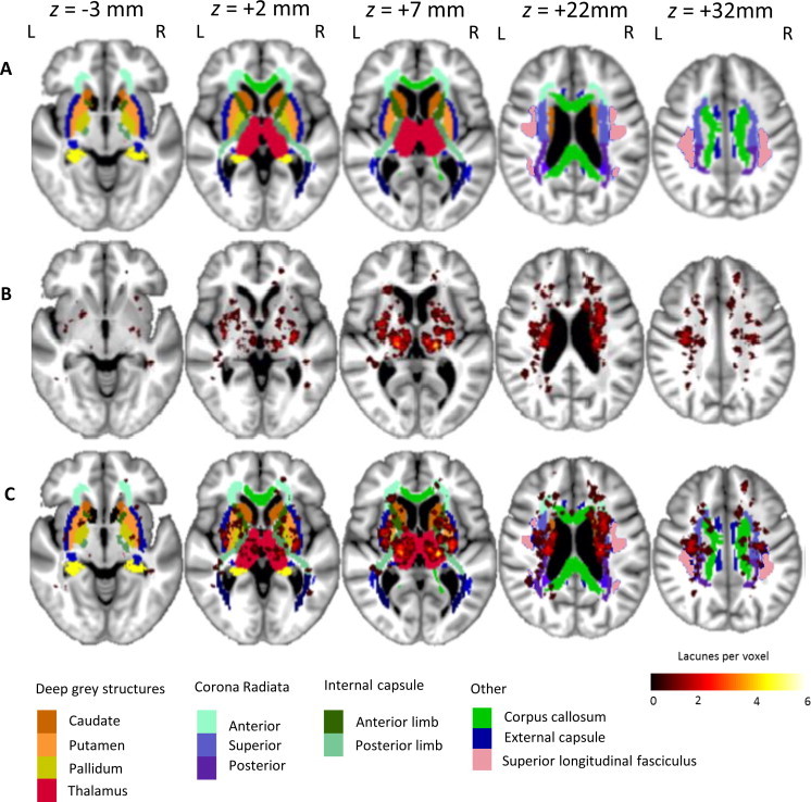
Objectives: Lacunes are an important disease feature of cerebral small vessel disease (SVD) but their relationship to cognitive impairment is not fully understood. To investigate this we determined (1) the relationship between lacune count and total lacune volume with cognition, (2) the spatial distribution of lacunes and the cognitive impact of lacune location, and (3) the whole brain anatomical covariance associated with these strategically located regions of lacune damage.

Methods: One hundred and twenty one patients with symptomatic lacunar stroke and radiological leukoaraiosis were recruited and multimodal MRI and neuropsychological data acquired. Lacunes were mapped semi-automatically and their volume calculated. Lacune location was automatically determined by projection onto atlases, including an atlas which segments the thalamus based on its connectivity to the cortex. Lacune locations were correlated with neuropsychological results. Voxel based morphometry was used to create anatomical covariance maps for these 'strategic' regions.

Results: Lacune number and lacune volume were positively associated with worse executive function (number p < 0.001; volume p < 0.001) and processing speed (number p < 0.001; volume p < 0.001). Thalamic lacunes, particularly those in regions with connectivity to the prefrontal cortex, were associated with impaired processing speed (Bonferroni corrected p = 0.016). Regions of associated anatomical covariance included the medial prefrontal, orbitofrontal, anterior insular cortex and the striatum.

Conclusion: Lacunes are important predictors of cognitive impairment in SVD. We highlight the importance of spatial distribution, particularly of anteromedial thalamic lacunes which are associated with impaired information processing speed and may mediate cognitive impairment via disruption of connectivity to the prefrontal cortex.

Keywords: Cognitive Impairment; Lacunes; Small vessel disease.


Figures

Fig. 1
Fig. 1
Lacune lesion location map showing spatial distribution throughout the brain. The map is superimposed onto the group-average T1-weighted 1 mm isotropic template and shown using the neurological viewing convention. Row A — atlas brain regions illustrating subcortical structures and white matter regions (standard atlases provided by the FSL package). A colour key to atlas regions is shown at the bottom of the figure. Row B — distribution of lacunes in MNI space. The colour bar at the bottom right indicates the number of lacunes present at each voxel. Row C — lacunes shown superimposed on atlas locations.
Fig. 2
Fig. 2
Oxford thalamic connectivity probability atlas superimposed on to group-average T1-weighted 1 mm isotropic template and shown using the neurological viewing convention. The colour key at the bottom of the figure represents the classification of thalamic voxels according to the cortical region with which they are connected with highest probability. Row A — thalamic regions according to the cortical region with which they are connected with highest probability. The colour key at the right of the figure represents this classification. Row B — lacunes shown superimposed on thalamic atlas locations. The colour bar on the right indicates number of lacunes present at each voxel. Row C — axial thalamic section from a cytoarchitectonic atlas (Morel et al., 1997). The nuclei have been coloured according to their major cortical connection site.
Fig. 3
Fig. 3
VBM analysis showing anatomical covariance maps of grey matter regions that positively co-varied with the prefrontal lacunar ROI average grey matter density. Statistical maps are shown superimposed on to the common group-average 1 mm isotropic template. The colour bar represents the t-statistic. Images are displayed in axial (row 1), sagittal (row 2) and coronal (row 3) orientations using the neurological viewing convention.
Fig. 4
Fig. 4
VBM analysis showing anatomical covariance maps of white matter regions that positively co-varied with prefrontal lacunar ROI average grey matter density. Statistical maps are shown superimposed on to the common group-average 1 mm isotropic template. The colour bar represents the t-statistic. Images are displayed in axial (row 1), sagittal (row 2) and coronal (row 3) orientations using the neurological viewing convention.

References

    1. Alexander-Bloch A., Giedd J., Bullmore E. Imaging structural co-variance between human brain regions. Nature Reviews. Neuroscience. 2013;14:322–336. 23531697 - PMC - PubMed
    1. Alexander G.E., DeLong M.R., Strick P.L. Parallel organization of functionally segregated circuits linking basal ganglia and cortex. Annual Review of Neuroscience. 1986;9:357–381. 3085570 - PubMed
    1. Ashburner J., Friston K.J. Diffeomorphic registration using geodesic shooting and Gauss–Newton optimisation. Neuroimage. 2011;55:954–967. 21216294 - PMC - PubMed
    1. Avants B., Tustison N., Song G., Cook P., Klein A., Gee J. A reproducible evaluation of ANTs similarity metric performance in brain image registration. Neuroimage. 2011;54:2033–2044. 20851191 - PMC - PubMed
    1. Bamford J., Sandercock P., Jones L., Warlow C. The natural history of lacunar infarction: the Oxfordshire Community Stroke Project. Stroke; a Journal of Cerebral Circulation. 1987;18:545–551. 3590244 - PubMed

Publication types