Review
. 2014 Jan;33(1):3-10.
doi: 10.14366/usg.13012. Epub 2013 Nov 26.

Practice guideline for the performance of breast ultrasound elastography

Affiliations
Review

Practice guideline for the performance of breast ultrasound elastography

Su Hyun Lee et al. Ultrasonography. 2014 Jan.

Abstract

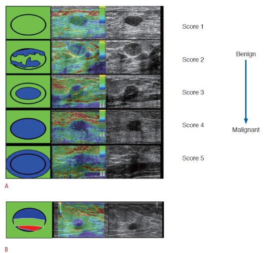
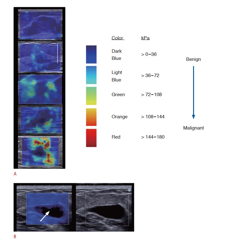
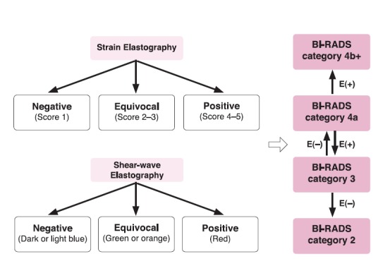
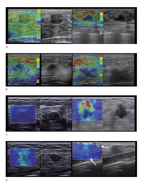
Ultrasound (US) elastography is a valuable imaging technique for tissue characterization. Two main types of elastography, strain and shear-wave, are commonly used to image breast tissue. The use of elastography is expected to increase, particularly with the increased use of US for breast screening. Recently, the US elastographic features of breast masses have been incorporated into the 2nd edition of the Breast Imaging Reporting and Data System (BI-RADS) US lexicon as associated findings. This review suggests practical guidelines for breast US elastography in consensus with the Korean Breast Elastography Study Group, which was formed in August 2013 to perform a multicenter prospective study on the use of elastography for US breast screening. This article is focused on the role of elastography in combination with B-mode US for the evaluation of breast masses. Practical tips for adequate data acquisition and the interpretation of elastography results are also presented.

Keywords: Breast, neoplasms; Elasticity imaging techniques; Ultrasonography.


Figures

Fig. 1.
Fig. 1.. Strain elastographic images of breast masses.
A. Five-point scale elasticity scores (Tsukuba score) with increasing probability of malignancy. A score of 1 indicates even strain throughout the entire hypoechoic lesion; a score of 2 indicates strain in most of the hypoechoic lesion with some areas of no strain; a score of 3 indicates strain at the periphery of the hypoechoic lesion with sparing of the center of the lesion; a score of 4 indicates no strain throughout the entire hypoechoic lesion; and a score of 5 indicates no strain throughout the entire hypoechoic lesion or in the surrounding area. B. An aliasing artifact that appears as a blue-green-red (BGR) pattern can be seen in a simple cyst. Note that a color code of red indicates soft, and blue is hard.
Fig. 2.
Fig. 2.. Shear-wave elastographic images of breast masses.
A. The ranges of maximum elasticity value according to the maximum elasticity color of breast masses are shown using a default color scale that ranges from 0 to +180 kPa. The maximum elasticity colors on shear-wave elastography (SWE) can be classified into three categories: dark blue and light blue indicating soft elasticity, green and orange indicating intermediate elasticity, and red indicating hard elasticity. B. A signal-void area (arrow) appears in a simple cyst.
Fig. 3.
Fig. 3.. A flowchart showing the combination of B-mode and elastographic results for the evaluation of breast masses.
Positive or negative elastographic results acquired on either strain elastography (SE) or shearwave elastography (SWE) can change the Breast Imaging Reporting and Data System (BI-RADS) category of breast masses. For the breast masses that present equivocal elastographic results, the B-mode ultrasound findings should be considered preferentially. E(+), positive result on SE or SWE; E(-), negative result on SE or SWE.
Fig. 4.
Fig. 4.. Representative good and poor quality elastographic images.
A. Good quality images on SE can be defined by a high signal-to-noise ratio in the region of interest. B. Poor quality images were acquired due to probe slipping (left) and a deeply located lesion or thick breast (right) on strain elastography. C. Good quality images with the typical appearance of a benign (left) and malignant mass (right) on shear-wave elastography (SWE). D. A benign mass can exhibit increased stiffness when the SWE image was acquired using compression (left). Artifacts can appear on SWE images (right) in the skin and chest wall (arrows).

References

    1. Itoh A, Ueno E, Tohno E, Kamma H, Takahashi H, Shiina T, et al. Breast disease: clinical application of US elastography for diagnosis. Radiology. 2006;239:341–350. - PubMed
    1. Cho N, Moon WK, Park JS, Cha JH, Jang M, Seong MH. Nonpalpable breast masses: evaluation by US elastography. Korean J Radiol. 2008;9:111–118. - PMC - PubMed
    1. Cho N, Jang M, Lyou CY, Park JS, Choi HY, Moon WK. Distinguishing benign from malignant masses at breast US: combined US elastography and color doppler US--influence on radiologist accuracy. Radiology. 2012;262:80–90. - PubMed
    1. Yi A, Cho N, Chang JM, Koo HR, La Yun B, Moon WK. Sonoelastography for 1,786 non-palpable breast masses: diagnostic value in the decision to biopsy. Eur Radiol. 2012;22:1033–1040. - PubMed
    1. Harvey J. Breast US: What's new in BI-RADS 2012? In: The 44th Annual Congress of Korean Society of Ultrasound in Medicine. In: The 44th Annual Congress of Korean Society of Ultrasound in Medicine; 2013 May 25; Seoul, Korea. 2013. p.138

LinkOut - more resources