. 2015 Oct;74(10):1924-30.
doi: 10.1136/annrheumdis-2014-205234. Epub 2014 Jun 16.

Preclinical characterisation of the GM-CSF receptor as a therapeutic target in rheumatoid arthritis

Affiliations

Preclinical characterisation of the GM-CSF receptor as a therapeutic target in rheumatoid arthritis

D E A Greven et al. Ann Rheum Dis. 2015 Oct.

Abstract

Objective: Previous work has suggested that the granulocyte macrophage colony stimulating factor (GM-CSF)-GM-CSF receptor α axis (GM-CSFRα) may provide a new therapeutic target for the treatment of rheumatoid arthritis (RA). Therefore, we investigated the cellular expression of GM-CSFRα in RA synovial tissue and investigated the effects of anti-GM-CSFRα antibody treatment in vitro and in vivo in a preclinical model of RA.

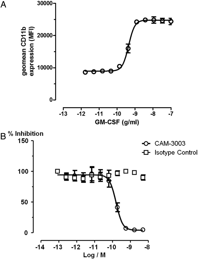
Methods: We compared GM-CSFRα expression on macrophages positive for CD68 or CD163 on synovial biopsy samples from patients with RA or psoriatic arthritis (PsA) to disease controls. In addition, we studied the effects of CAM-3003, an anti-GM-CSFR antibody in a collagen induced arthritis model of RA in DBA/1 mice. The pharmacokinetic profile of CAM-3003 was studied in naïve CD1(ICR) mice (see online supplement) and used to interpret the results of the pharmacodynamic studies in BALB/c mice.

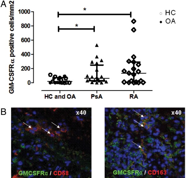
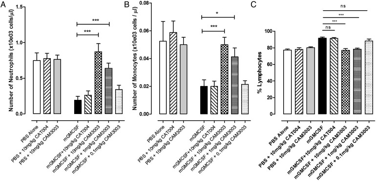
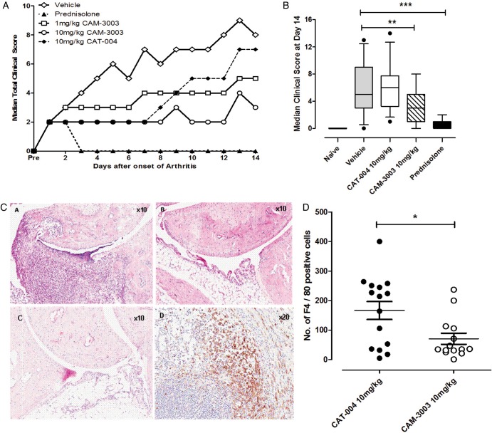
Results: GM-CSFRα was expressed by CD68 positive and CD163 positive macrophages in the synovium, and there was a significant increase in GM-CSFRα positive cells in patients in patients with RA as well as patients with PsA compared with patients with osteoarthritis and healthy controls. In the collagen induced arthritis model there was a dose dependent reduction of clinical arthritis scores and the number of F4/80 positive macrophages in the inflamed synovium after CAM-3003 treatment. In BALB/c mice CAM-3003 inhibited recombinant GM-CSF mediated margination of peripheral blood monocytes and neutrophils.

Conclusions: The findings support the ongoing development of therapies aimed at interfering with GM-CSF or its receptor in various forms of arthritis, such as RA and PsA.

Keywords: Pharmacokinetics; Rheumatoid Arthritis; Treatment.


Figures

Figure 1
Figure 1
(A) The quantification of granulocyte macrophage colony stimulating factor (GM-CSFRα) positive cells in ST from patients with rheumatoid arthritis (RA) and psoriatic arthritis (PsA) (inflammatory arthritis; n=50) compared with ST from patients with osteoarthritis (OA) and healthy controls (HCs) (non-inflammatory; n=19). Median (IQRs) are shown. *p Value <0.05 (Mann–Whitney U test). (B) Representative picture of an immunofluorescence staining for GM-CSFRα (green). GM-CSFRα shows colocalisation with either CD68 (red) or CD163 (red) (arrows).
Figure 2
Figure 2
Characterisation of the cell surface expression of CD11b on mouse granulocytes stimulated with mouse granulocyte macrophage colony stimulating factor (GM-CSF). Mouse granulocytes were stimulated with a dose response of mouse GM-CSF for 1 hr and cell surface expression of CD11b quantified by flow cytometry using the mean fluorescent intensity (MFI). GM-CSF was able to dose dependently increase CD11b expression. (B) Mouse granulocytes were stimulated with mouse GM-CSF corresponding to the EC80 (2.5 ng/mL) and incubated with increasing concentrations of CAM-3003 for 1.5 h. Cells were then labelled for CD11b expression and the MFI quantified by flow cytometry. CAM-3003 dose-dependently inhibited GM-CSF induced CD11b upregulation.
Figure 3
Figure 3
(A) Characterisation of the in vivo neutralising activity of CAM-3003 in a model of granulocyte macrophage colony stimulating factor (GM-CSF) induced leucocyte margination. BALB/c mice were dosed intravenously with either CAM-3003 at 10 m/kg, 1 m/kg and 0.1 m/kg, the isotype control (CAT-004) at 10 mg/kg or vehicle (PBS) and then stimulated with mouse GM-CSF (mGMCSF, 0.25 μg) for 15 min. Peripheral blood was analysed using an ADVIA blood cell counter. (A) Absolute number of circulating neutrophils. (B) Absolute number of circulating monocytes. (C) Percentage of circulating lymphocytes 15 min post administration of GM-CSF. Statistically significant changes were observed due to changes in percentage of neutrophils and monocytes due to margination. Absolute numbers of lymphocytes did not change (data not shown). Data are expressed as mean±SEM (n=7–8). *p<0.05; ***p<0.001.
Figure 4
Figure 4
(A) Characterisation of the effect of GM-CSFRα inhibition in established arthritis. (A) Mice were treated daily for 14 days post the onset of arthritis with either CAM-3003 at 10 mg/kg or 1 mg/kg, CAT-004 (10 mg/kg) as an isotype control, prednisolone (dose) or vehicle alone. Median clinical score was plotted daily to map disease progression. (B) Comparison of mean clinical score at Day 14 from three independent studies. Mean time of onset of arthritis was 28±7 days. Data is expressed as mean±SEM. (C) Histological images of mouse ankle joint from collagen induced arthritis model. Panel A–C are representative H&E images of (A) CIA induced arthritic joint treated with an isotype control antibody (CAT-004; 10mg/kg), (B) CIA induced arthritic joint treated with anti-GM-CSFRα antibody (CAM-3003; 10 mg/kg) (C) naive mouse joint (D) immunohistochemistry staining of a CIA induced arthritic mouse joint, demonstrating an infiltration of F4/80+ cells into the synovium. (D) Quantification of total numbers of F4/80+ cells located within the annotated area (see online supplementary figure S2) within the synovial membrane area. Data are from a collagen induced arthritis model treated with either an antimurine GM-CSFRα antibody (CAM-3003) or an isotype control antibody (CAT-004). Macrophages were assessed by positive membrane staining with anti-F4/80 immunohistochemistry. Data shown represent total F4/80+ counts for both hind paws of animals (n=14/15 per group) and are represented as mean±SEM. *p<0.05.

References

    1. Hamilton JA, Tak PP. The dynamics of macrophage lineage populations in inflammatory and autoimmune diseases. Arthritis Rheum 2009;60:1210–21. - PubMed
    1. van Nieuwenhuijze A, Koenders M, Roeleveld D, et al. GM-CSF as a therapeutic target in inflammatory diseases. Mol Immunol 2013;56:675–82. - PubMed
    1. Cornish AL, Campbell IK, McKenzie BS, et al. G-CSF and GM-CSF as therapeutic targets in rheumatoid arthritis. Nat Rev Rheumatol 2009;5:554–9. - PubMed
    1. Hamilton JA. Colony-stimulating factors in inflammation and autoimmunity. Nat Rev Immunol 2008;8:533–44. - PubMed
    1. Hamilton JA, Anderson GP. GM-CSF Biology. Growth Factors 2004;22:225–31. - PubMed

Publication types

MeSH terms

Substances