. 2014 Jun 17:4:5290.
doi: 10.1038/srep05290.

Efficient genetic manipulation of the NOD-Rag1-/-IL2RgammaC-null mouse by combining in vitro fertilization and CRISPR/Cas9 technology

Affiliations

Efficient genetic manipulation of the NOD-Rag1-/-IL2RgammaC-null mouse by combining in vitro fertilization and CRISPR/Cas9 technology

Feng Li et al. Sci Rep. .

Abstract

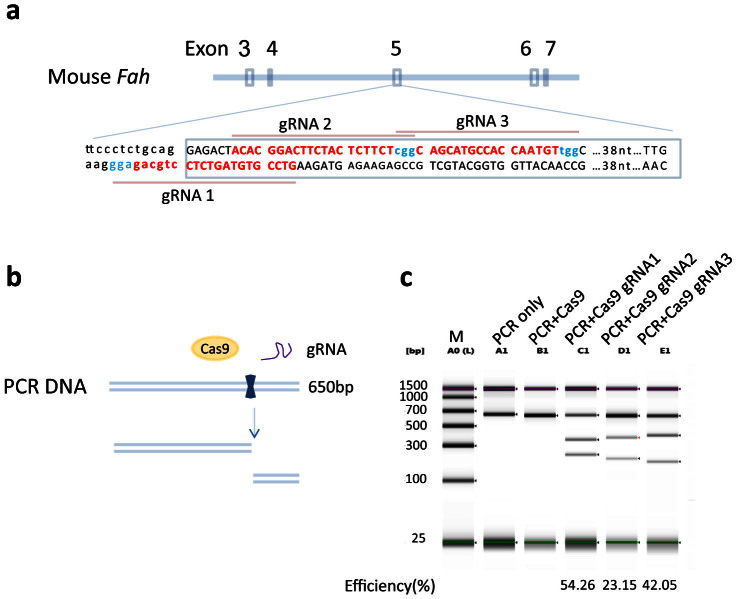
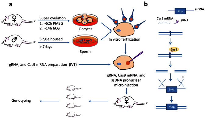
Humanized mouse models have become increasingly important and widely used in modeling human diseases in biomedical research. Immunodeficient mice such as NOD-Rag1-/-IL2RgammaC-null (NRG) or NOD-SCID-IL2RgammaC-null (NSG) mice are critical for efficient engraftment of human cells or tissues. However, their genetic modification remains challenging due to a lack of embryonic stem cells and difficulty in the collection of timed embryos after superovulation. Here, we report the generation of gene knockout NRG mice by combining in vitro fertilization (IVF) and CRISPR/Cas9 technology. Sufficient numbers of fertilized embryos were produced through IVF, and a high rate of Fah gene targeting was achieved with microinjection of Cas9 mRNA, gRNA and single strand oligonucleotide DNA (ssDNA) into the embryos. The technology paves the way to construct NRG or NSG mutant mice to facilitate new humanized mouse models. The technology can also be readily adapted to introduce mutations in other species such as swine and non-human primates.


Conflict of interest statement

Yes, there is potential Competing Interest. D. Cowley is employed by, has equity ownership in and serves on the board of directors of TransViragen, the company which has been contracted by UNC-Chapel Hill to manage its Animal Models Core Facility. No other authors have any competing financial interests.

Figures

Figure 1
Figure 1. gRNAs targeting Fah exon5 can efficiently direct Cas9 cleavage of target DNA.
(a) gRNA sequences against Fah exon5. The exon 3, 4, 5 and 6 region of the mouse Fah gene is shown. The exon5 sequence (upper case) and part of the upstream intron sequence (lower case) are shown with 3 gRNA sequences (labeled in red), and the PAM sequence NGGs in turquoise. (b) and (c) gRNA cleavage efficiency in vitro. (b) Schematic diagram of gRNA mediated Cas9 DNA cleavage. The PCR DNA product containing the gRNA target sites, Cas9 nuclease protein and gRNA are mixed. The Cas9/gRNA complex cuts the PCR product into two fragments. (c) The cleaved DNA was resolved on TapeStation. A0(L), DNA ladder; A1, PCR DNA alone; B1, PCR DNA + Cas9 nuclease; C1, D1 and E1, PCR DNA + Cas9 nuclease + gRNA1 (C1), gRNA2 (D1) and gRNA 3 (E1). Full-length gel is shown. The gRNA mediated cutting efficiency is shown below each lane.
Figure 2
Figure 2. Strategy to generate NRG Fah knockout mice.
(a) Schematic illustration of IVF and pronuclear microinjection. Female NRG mice are superovulated with PMSG and hCG followed by oocyte collection. Sperm is collected from male NRG mice. The oocytes and sperm are incubated to generate fertilized eggs and embryos, which are then microinjected with gRNA, Cas9 mRNA and ssDNA in the pronucleus. The injected embryos are then transferred into pseudopregnant surrogate mothers. Mouse pups are genotyped. (b) Mechanism of gRNA, Cas9 mRNA, and ssDNA mediated Fah gene knockout. Cas9 mRNA is translated into Cas9 protein after microinjection. The Cas9/gRNA complex binds the genomic DNA and generates DSBs. The ssDNA contains homologous sequence spanning the double strand break sites with ~50 bp on each side, two stop codons and a BamHI site. The ssDNA can be used as a template for homologous recombination to introduce the stop codons and BamHI site. The mouse is drawn by the authors (F.L. and L.S.), using Adobe Photoshop and Adobe Illustrator.
Figure 3
Figure 3. Genotyping and sequence analysis of founder mice.
(a) PCR results of founder mice. Mice from two microinjection sessions were genotyped by PCR amplification and BamHI digestion. PCR and BamHI digestion products were electrophoresed in 1.2% argorose gel. Mice from the first injection session (wild type Cas9 mRNA) were labeled #1-1 to #1-10, and mice from the second injection session (mutant D10A Cas9 mRNA) were labeled #2-1 to #2-10. Wild type mouse genomic DNA was used as negative control (WT con). Cropped gels are shown. Full-length gels are provided for review in the supplementary file. (b) Sequence analysis of the mutated Fah alleles. The wild type sequence of exon5 is shown on top, with the encoded amino acids indicated above the sequence. The gRNA targeting sites are shown in red letters and PAM NGGs in turquoise. The mutant alleles of each mouse are labeled with A and B, following the mouse ID number. The indels for each mutated allele from different mice are shown in the middle. The deleted sequences are marked in gray, and the inserted sequences are shown below the wild type sequence. The deletion or insertion length of each indel is shown on the right. The Fah mutant alleles with the knock-in sequence are shown in the lower panel. Only one sequence is shown for the stop codon (@) and BamHI insertion. Indel, insertion and deletion; in, insertion; del, deletion.
Figure 4
Figure 4. Germline transmission of the Fah mutant allele.
(a) Primers designed to genotype the Fah mutant allele by PCR. Primers FAH-F1, FAH-R1 and FAH-probe are used in the same PCR reaction. Primers FAH-F1 and FAH-R1 amplify a 560 bp segment from the WT and 565 bp from the STOP-BamHI knock-in allele. The primers FAH-F1 and FAH-probe, specifically targeting the knock-in sequence, only amplify a 454 bp segment. (b) PCR genotyping results of F1 generation mice. The mouse numbers, from F0001 to F0010, are shown above each lane. Founder #1-1 and wild type mouse genomic DNA serve as positive and negative controls, respectively. Blank is a reaction without any DNA template. The amplicon size is shown on the right. The gender of each mouse is shown below each lane. ♀, female; ♂, male. Left lane, DNA ladder. Cropped gel is shown. Full-length gel is provided for review in the supplementary file.

References

    1. Zhang L. & Su L. HIV-1 immunopathogenesis in humanized mouse models. Cell Mol Immunol 9, 237–244 (2012). - PMC - PubMed
    1. Brehm M. A., Jouvet N., Greiner D. L. & Shultz L. D. Humanized mice for the study of infectious diseases. Curr Opin Immunol 25, 428–435 (2013). - PMC - PubMed
    1. Brehm M. A. & Shultz L. D. Human allograft rejection in humanized mice: a historical perspective. Cell Mol Immunol 9, 225–231 (2012). - PMC - PubMed
    1. Takebe T. et al. Vascularized and functional human liver from an iPSC-derived organ bud transplant. Nature 499, 481–484 (2013). - PubMed
    1. Zhu S. et al. Mouse liver repopulation with hepatocytes generated from human fibroblasts. Nature 508, 93–97 (2014). - PMC - PubMed

Publication types

MeSH terms