

Review
doi: 10.1259/bjr.20140216.
Epub 2014 Jun 17.
Imaging of acute stroke prior to treatment: current practice and evolving techniques
Affiliations
- PMID: 24936980
- PMCID: PMC4112390
- DOI: 10.1259/bjr.20140216
Item in Clipboard
Review
Imaging of acute stroke prior to treatment: current practice and evolving techniques
Br J Radiol.
2014 Aug.
Abstract
Standard imaging in acute stroke is undertaken with the aim of diagnosing the underlying cause and excluding stroke mimics. In the presence of ischaemic stroke, imaging is also needed to assess patient suitability for treatment with intravenous thrombolysis. Non-contrast CT is predominantly used, but MRI can also exclude any contraindications to thrombolysis treatment. Advanced stroke imaging such as CT and MR angiography and perfusion imaging are increasingly used in an acute setting. In this review, we discuss the evidence for the application of these advanced techniques in the imaging of acute stroke.
Figures

Acute stroke changes on non-contrast CT. Left image shows an area of hypodensity and loss of grey–white matter differentiation affecting the right basal ganglia and insular cortex (within box) consistent with an acute infarct. Compare with the normal left side. Right image shows a hyperdense mainstem of left middle cerebral artery in a different patient (arrow).

Acute stroke changes on MRI. Acute lacunar infarct within the left thalamus as demonstrated on MRI. Infarct is high signal on T2 weighted (left) and diffusion-weighted imaging (middle) but correspondingly low signal on apparent diffusion coefficient imaging (right).
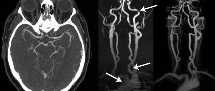
CT and MR angiography. Left image shows normal CT angiography through the circle of Willis. Time-of-flight MR angiography (middle image) and contrast-enhanced MR angiography (right image) of the neck arteries are compared in the coronal plane. Note significant artefacts on time-of-flight images (arrows).

CT angiography (CTA) source images. CTA (left image) demonstrates an occlusion within the proximal left middle cerebral artery (arrow). When window settings are adjusted to view brain (right image), these “CTA source images” reveal that the left middle cerebral artery territory (within dotted line) is underperfused; affected area is hypodense relative to contralateral normal brain.

CT perfusion maps showing commonly provided perfusion parameters of cerebral blood flow (CBF), cerebral blood volume (CBV), mean transit time (MTT) and time to peak (TTP). Anterior left middle cerebral artery territory perfusion defect showing decreased CBF and increased MTT and TTP. CBV is marginally increased. The difference between CBV and the other parameters suggests ischaemic but not yet infarcted tissue (i.e. penumbra, at risk). This patient proceeded to treatment with intravenous thrombolysis. P, posterior; R, right.
References
-
- Mathers CD, Bernand C, Iburg KM, Inoue M, Ma Fat D, Shibuya K, et al. . Global burden of disease: data sources, methods and results. World Health Organization; 2004. [Cited 30 June 2014.] Available from: http://www.who.int/healthinfo/global_burden_disease/en/
-
- The International Stroke Trial (IST): a randomised trial of aspirin, subcutaneous heparin, both, or neither among 19435 patients with acute ischaemic stroke. International Stroke Trial Collaborative Group. Lancet 1997; 349: 1569–81. - PubMed
-
- Dombrowski SU, Mackintosh JE, Sniehotta FF, Araujo-Soares V, Rodgers H, Thomson RG, et al. . The impact of the UK “Act FAST” stroke awareness campaign: content analysis of patients, witness and primary care clinicians' perceptions. BMC Public Health 2013; 13: 915. doi: 10.1186/1471-2458-13-915 - DOI - PMC - PubMed
Publication types
MeSH terms
Grants and funding
LinkOut - more resources
Full Text Sources
Other Literature Sources
Medical
Molecular Biology Databases
