

SPF rabbits infected with rabbit hepatitis E virus isolate experimentally showing the chronicity of hepatitis
- PMID: 24937350
- PMCID: PMC4061063
- DOI: 10.1371/journal.pone.0099861
SPF rabbits infected with rabbit hepatitis E virus isolate experimentally showing the chronicity of hepatitis
Abstract
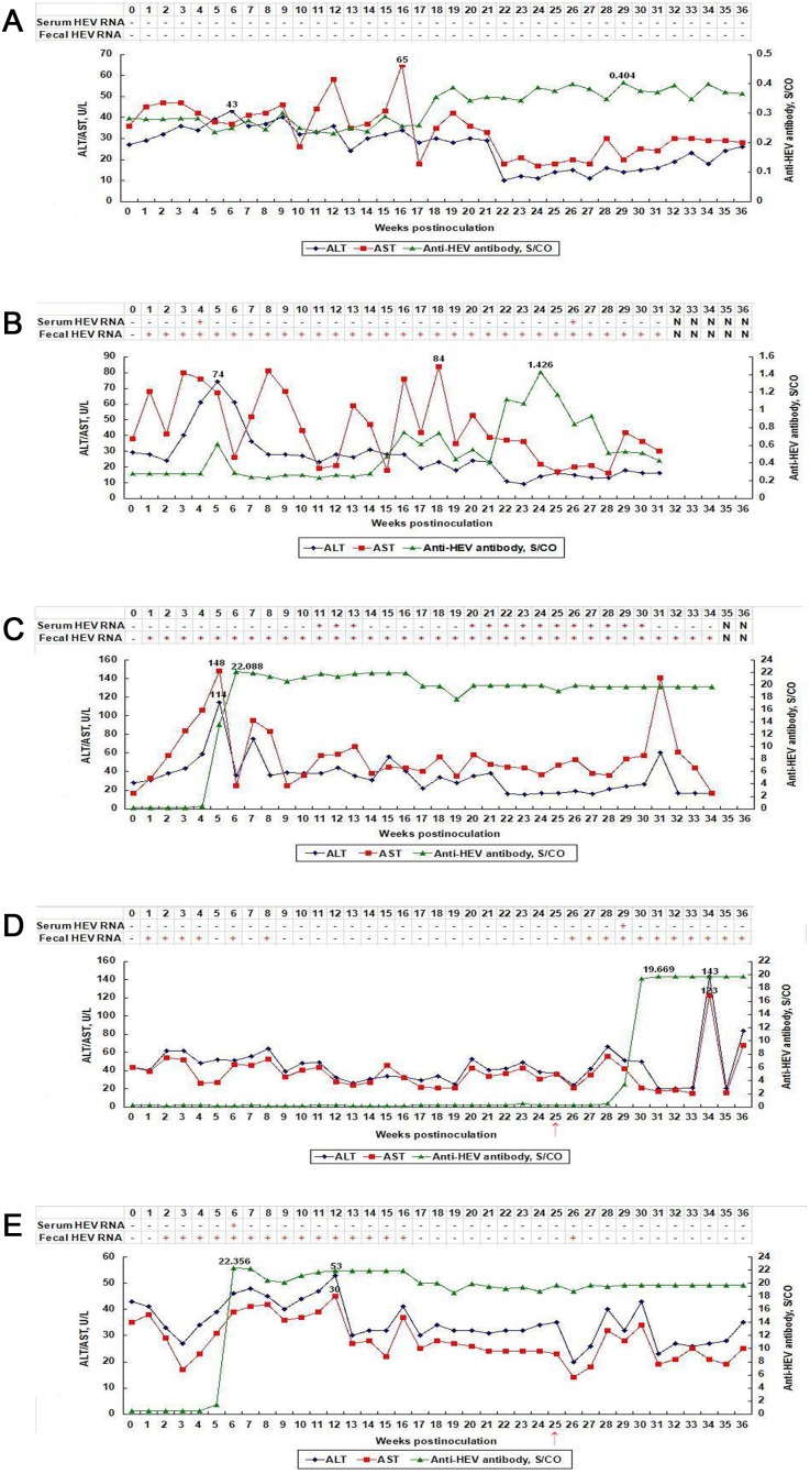
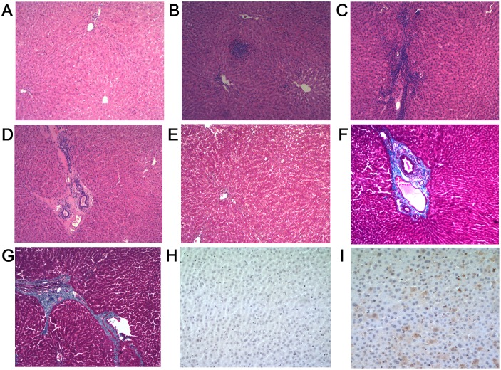
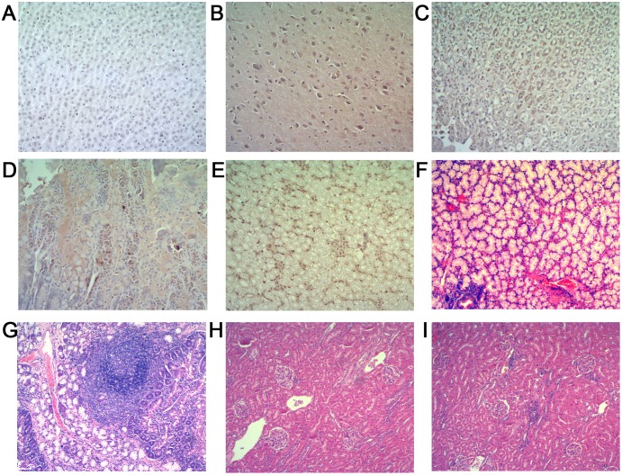
This study focused on investigating the pathogenesis seen in specific-pathogen-free (SPF) rabbits following infection with a homologous rabbit HEV isolate (CHN-BJ-rb14) and comparing it to that seen following infection with a heterologous swine genotype 4 HEV isolate (CHN-XJ-SW13). Three of the four animals inoculated with the homologous rabbit HEV became infected, exhibiting an intermittent viremia, obvious fluctuations of liver function biomarkers alanine aminotransferase (ALT) and aspartate aminotransferase (AST), and persistent fecal virus shedding throughout the nine month study. In addition, liver histopathology showed both chronic inflammation and some degree of fibrosis. Both positive and negative-stranded HEV RNA and HEV antigen expression were detected in liver, brain, stomach, duodenum and kidney from the necropsied rabbits. Inflammation of extrahepatic tissue (duodenum and kidney) was also observed. Three of the four rabbits inoculated with the heterologous genotype 4 swine HEV also became infected, showing similar levels of anti-HEV antibody to that generated following infection with the homologous virus isolate. The duration of both viremia and fecal shedding of virus was however shorter following infection with the heterologous virus and there was no significant elevation of liver function biomarkers. These results suggest that rabbit HEV infection may cause more severe hepatitis and prolong the course of the disease, with a possible chronic trend of hepatitis in SPF rabbits.
Conflict of interest statement
Figures
Similar articles
-
Different susceptibility and pathogenesis of rabbit genotype 3 hepatitis E virus (HEV-3) and human HEV-3 (JRC-HE3) in SPF rabbits.Vet Microbiol. 2017 Aug;207:1-6. doi: 10.1016/j.vetmic.2017.05.019. Epub 2017 May 29. Vet Microbiol. 2017. PMID: 28757007
-
Experimental infection of rabbits with rabbit and genotypes 1 and 4 hepatitis E viruses.PLoS One. 2010 Feb 11;5(2):e9160. doi: 10.1371/journal.pone.0009160. PLoS One. 2010. PMID: 20161794 Free PMC article.
-
Cross-species infection of mice by rabbit hepatitis E virus.Vet Microbiol. 2018 Nov;225:48-52. doi: 10.1016/j.vetmic.2018.09.015. Epub 2018 Sep 18. Vet Microbiol. 2018. PMID: 30322532
-
An overview: Rabbit hepatitis E virus (HEV) and rabbit providing an animal model for HEV study.Rev Med Virol. 2018 Jan;28(1). doi: 10.1002/rmv.1961. Epub 2017 Nov 17. Rev Med Virol. 2018. PMID: 29148605 Review.
-
Advances in Hepatitis E Virus Biology and Pathogenesis.Viruses. 2021 Feb 9;13(2):267. doi: 10.3390/v13020267. Viruses. 2021. PMID: 33572257 Free PMC article. Review.
Cited by
-
Experimental infection of hepatitis E virus induces pancreatic necroptosis in miniature pigs.Sci Rep. 2020 Jul 21;10(1):12022. doi: 10.1038/s41598-020-68959-3. Sci Rep. 2020. PMID: 32694702 Free PMC article.
-
Hepatitis E virus immunosuppressed animal models.BMC Infect Dis. 2024 Sep 12;24(1):965. doi: 10.1186/s12879-024-09870-4. BMC Infect Dis. 2024. PMID: 39266958 Free PMC article. Review.
-
The Amino-Terminal Region of Hepatitis E Virus ORF1 Containing a Methyltransferase (Met) and a Papain-Like Cysteine Protease (PCP) Domain Counteracts Type I Interferon Response.Viruses. 2018 Dec 18;10(12):726. doi: 10.3390/v10120726. Viruses. 2018. PMID: 30567349 Free PMC article.
-
Hepatitis E Virus Quasispecies in Cerebrospinal Fluid with Neurological Manifestations.Vaccines (Basel). 2021 Oct 19;9(10):1205. doi: 10.3390/vaccines9101205. Vaccines (Basel). 2021. PMID: 34696313 Free PMC article.
-
Animal Models for Hepatitis E virus.Viruses. 2019 Jun 18;11(6):564. doi: 10.3390/v11060564. Viruses. 2019. PMID: 31216711 Free PMC article. Review.
References
-
- Kamar N, Bendall R, Legrand-Abravanel F, Xia NS, Ijaz S, et al. (2012) Hepatitis E. Lancet. 379: 2477–2488. - PubMed
-
- Kamar N, Selves J, Mansuy JM, Ouezzani L, Peron JM, et al. (2008) Hepatitis E virus and chronic hepatitis in organ-transplant recipients. N Engl J Med 358: 811–817. - PubMed
-
- Dalton HR, Bendall RP, Keane FE, Tedder RS, Ijaz S (2009) Persistent carriage of hepatitis E virus in patients with HIV infection. N Engl J Med 361: 1025–1027. - PubMed
-
- Tavitian S, Peron JM, Huynh A, Mansuy JM, Ysebaert L, et al. (2010) Hepatitis E virus excretion can be prolonged in patients with hematological malignancies. J Clin Virol 49: 141–144. - PubMed
-
- Khuroo MS (1980) Study of an epidemic of non-A, non-B hepatitis. Possibility of another human hepatitis virus distinct from post-transfusion non-A, non-B type. Am J Med 68: 818–824. - PubMed
Publication types
MeSH terms
Substances
LinkOut - more resources
Full Text Sources
Other Literature Sources
