. 2014 Aug;31(8):983-91.
doi: 10.1007/s10815-014-0271-7. Epub 2014 Jun 18.

Development of a high-yield technique to isolate spermatogonial stem cells from porcine testes

Affiliations

Development of a high-yield technique to isolate spermatogonial stem cells from porcine testes

Min Hee Park et al. J Assist Reprod Genet. 2014 Aug.

Abstract

Purpose: To date, the methods available for isolating spermatogonial stem cells (SSCs) from porcine testicular cells have a low efficiency of cell separating. Therefore, we tried to develop a novel isolation technique with a high-yield cell separating ability to isolate SSCs from porcine testes.

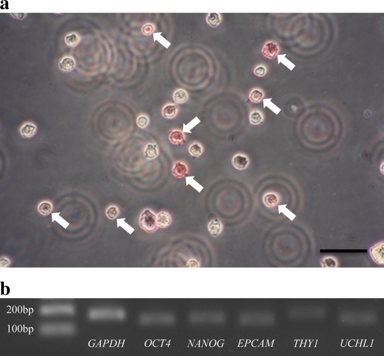
Methods: We confirmed the presence of SSCs by measuring alkaline phosphatase (AP) activity and SSC-specific gene expression in neonatal porcine testis-derived testicular cells. Subsequently, the isolation of SSCs from testicular cells was performed using different techniques as follows: differential plating (DP), double DP, Petri dish plating post-DP, magnetic-activated cell sorting (MACS), and MACS post-DP. Positive AP staining was used to assess and compare the isolation efficiency of each method.

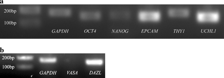
Results: Petri dish plating post-DP resulted in the highest isolation efficiency. The putative SSCs isolated using this method was then further characterized by analyzing the expression of SSC-specific genes and -related proteins, and germ cell-specific genes. OCT4, NANOG, EPCAM, THY1, and UCHL1 were expressed transcriptionally, and OCT4, NANOG, SOX2, TRA-1-60, TRA-1-81, and PLZF were expressed translationally in 86 % of the isolated SSCs. In contrast, no difference was observed in the percentage of cells expressing luteinizing hormone receptor (LHR), a Leydig cell-specific protein, or GATA4, a Sertoli cell-specific protein, between SSCs and negative control cells. In addition, transcriptional expression of VASA, a primordial germ cell-specific marker, and DAZL, a premeiotic germ cell-specific marker, wasn't and was detected, respectively.

Conclusions: We successfully developed a novel high-yield technique to isolate SSCs from porcine testes to facilitate future porcine SSC-related research.


Figures

Fig. 1
Fig. 1
Experimental design
Fig. 2
Fig. 2
Existence of SSCs in testicular cells derived from neonatal porcine testes. Testes derived from neonatal male porcine were enzymatically dissociated and the presence of SSCs in dissociated testicular cells was identified by conducting alkaline phosphatase (AP) staining and analysis of SSC-specific genes expression through reverse transcription-polymerase chain reaction (RT-PCR). As the results, there were observed testicular cells stained positively for AP activity (arrow, a) and showing the expression of OCT4 (151 bp), NANOG (158 bp), EPCAM (154 bp), THY1 (178 bp) and UCHL1 (150 bp) (b). GAPDH (180 bp) was used as an internal control. n = 3. Scale bars represent 200 μm
Fig. 3
Fig. 3
Comparison of isolation efficiency of SSCs from neonatal porcine testicular cells according to different SSC isolation techniques. Putative SSC populations were retrieved from testicular cells derived from neonatal porcine testes by each SSC isolation method and stained for alkaline phosphatase (AP) activity. The SSCs retrieved by Petri dish plating post-DP method showed significantly the highest percentage of SSCs stained positively by AP staining. All data shown are means ± SD of five independent experiments. *-**** p < 0.05. n = 3
Fig. 4
Fig. 4
Reverse transcription-polymerase chain reaction (RT-PCR) analysis for the expression of SSC- and germ cell-specific genes in porcine SSCs isolated by Petri dish plating post-DP method. Isolated SSCs showed successfully transcriptional expression of OCT4 (151 bp), NANOG (158 bp), EPCAM (154 bp), THY1 (178 bp) and UCHL1 (150 bp) (SSC-specific marker genes) (a), with DAZL (153 bp) (premeiotic germ cell-specific marker gene) (b). However, any transcripts derived from VASA (195 bp) (primordial germ cell-specific marker gene) weren’t detected (b). GAPDH (180 bp) was used as an internal control. n = 3
Fig. 5
Fig. 5
Translational expression of SSC-specific genes in porcine SSCs isolated by Petri dish plating post-DP method. The yield of SSCs stained positively with antibodies detecting SSC-specific proteins was measured by flow cytometry analysis. The expression of OCT4, NANOG, SOX2, TRA-1-60, TRA-1-81 and PLZF was identified in isolated SSCs (a) and at least 95 % or more of SSCs showed positivity against OCT4, NANOG, SOX2, and PLZF expression. Whereas, the expression of TRA-1-60 and TRA-1-81 was detected in at least 86 % or more of SSCs and the percentage of SSCs stained positively by TRA-1-60 and TRA-1-81 antibodies was significantly lower than those of the other markers. Moreover, localization of TRA-1-60 and TRA-1-81 on SSC surface membrane was identified by immunocytochemistry (b). All data shown are means ± SD of three independent experiments. Negative control means the unstained SSCs. Scale bar is 40 μm.*–*** p < 0.05
Fig. 6
Fig. 6
The percentage of Leydig and Sertoli cells coexisted in porcine SSCs isolated by Petri dish plating post-DP method. The isolated SSCs were stained with Leydig cell-specific LHR and Sertoli cell-specific GATA4 antibodies and the yield of Leydig and Sertoli cells were analyzed by flow cytometry. As the results, the percentage of cells showing positivity against LHR and GATA4 antibodies wasn’t significantly different from those of the unstained SSCs (negative control). All data shown are means ± SD of three independent experiments

Similar articles

Cited by

References

    1. McLaren A. Primordial germ cells in the mouse. Dev Biol. 2003;262(1):1–15. doi: 10.1016/S0012-1606(03)00214-8. - DOI - PubMed
    1. Brinster RL. Germline stem cell transplantation and transgenesis. Science. 2002;296:2174–2176. doi: 10.1126/science.1071607. - DOI - PMC - PubMed
    1. Yang Y, Honaramooz A. Efficient purification of neonatal porcine gonocytes with Nycodenz and differential plating. Reprod Fertil Dev. 2011;23:496–505. doi: 10.1071/RD10042. - DOI - PubMed
    1. Dym M. Spermatogonial stem cells of the testis. PNAS. 1994;91:11287–11289. doi: 10.1073/pnas.91.24.11287. - DOI - PMC - PubMed
    1. De Rooij DG. Stem cells in the testis. Int J Exp Pathol. 1998;79:67–80. doi: 10.1046/j.1365-2613.1998.00057.x. - DOI - PMC - PubMed

Publication types