. 2014 Jun 17:2:67.
doi: 10.1186/2051-5960-2-67.

Selective vulnerability of the cerebral vasculature to blast injury in a rat model of mild traumatic brain injury

Affiliations

Selective vulnerability of the cerebral vasculature to blast injury in a rat model of mild traumatic brain injury

Miguel A Gama Sosa et al. Acta Neuropathol Commun. .

Abstract

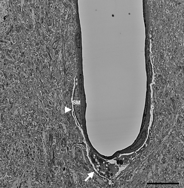
Background: Blast-related traumatic brain injury (TBI) is a common cause of injury in the military operations in Iraq and Afghanistan. How the primary blast wave affects the brain is not well understood. The aim of the present study was to examine whether blast exposure affects the cerebral vasculature in a rodent model. We analyzed the brains of rats exposed to single or multiple (three) 74.5 kPa blast exposures, conditions that mimic a mild TBI. Rats were sacrificed 24 hours or between 6 and 10 months after exposure. Blast-induced cerebral vascular pathology was examined by a combination of light microscopy, immunohistochemistry, and electron microscopy.

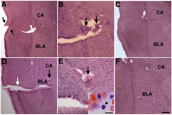
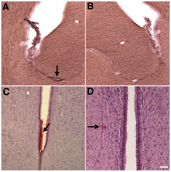
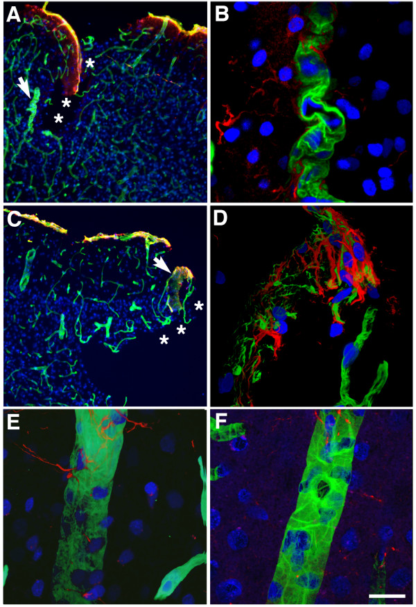
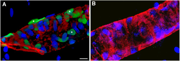
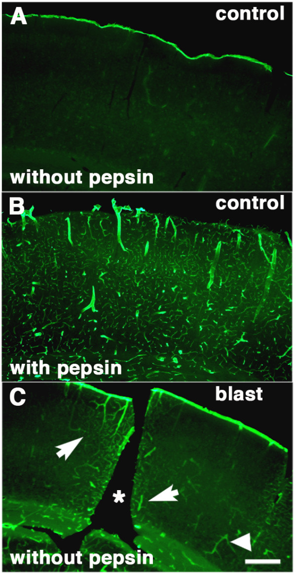
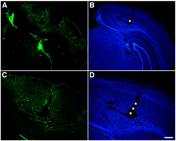
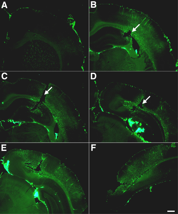
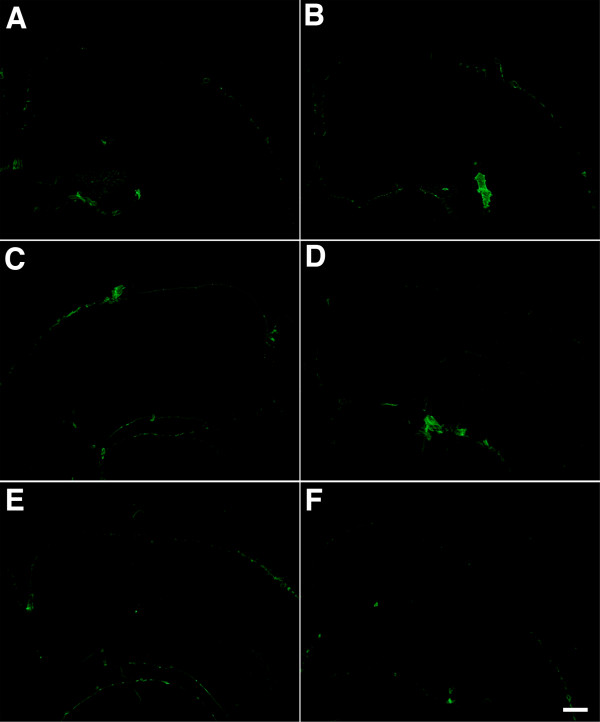
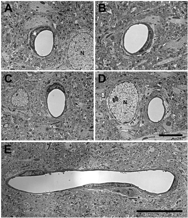
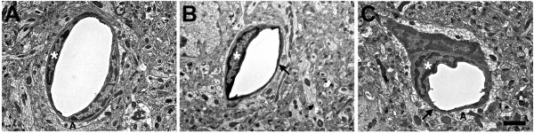
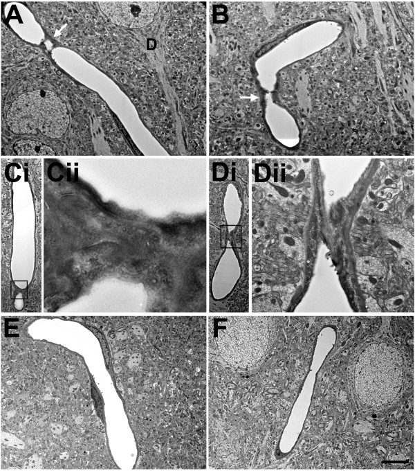
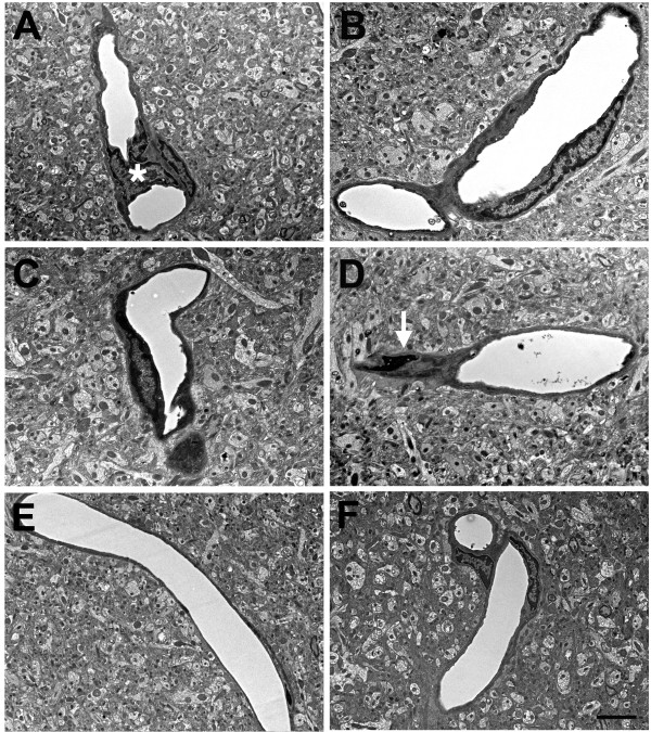
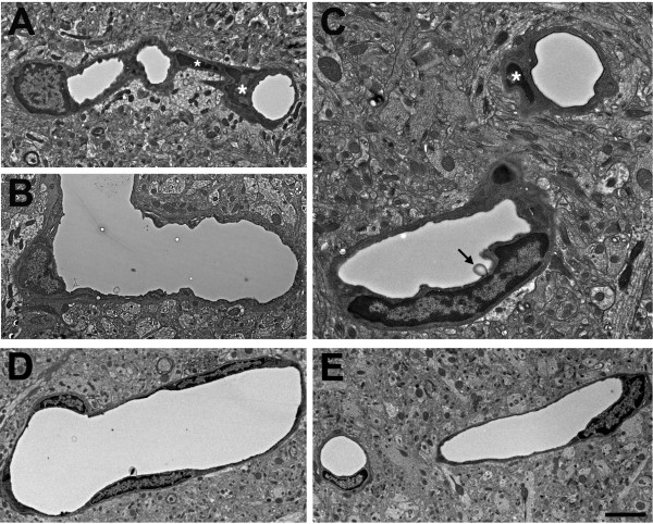
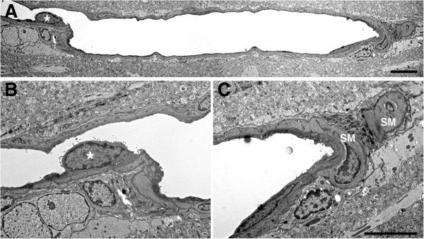
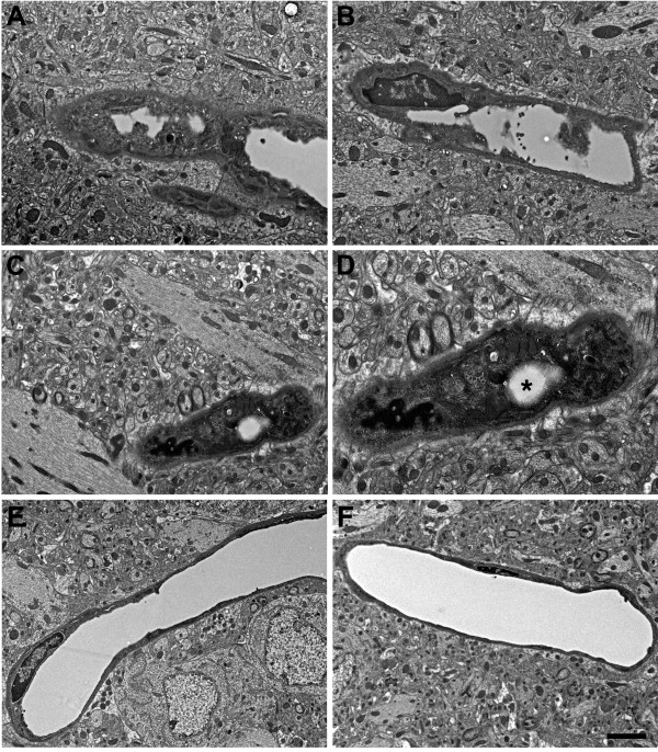
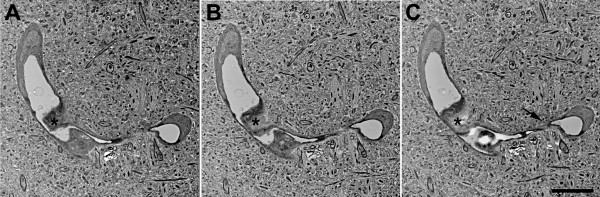
Results: We describe a selective vascular pathology that is present acutely at 24 hours after injury. The vascular pathology is found at the margins of focal shear-related injuries that, as we previously showed, typically follow the patterns of penetrating cortical vessels. However, changes in the microvasculature extend beyond the margins of such lesions. Electron microscopy revealed that microvascular pathology is found in regions of the brain with an otherwise normal neuropil. This initial injury leads to chronic changes in the microvasculature that are still evident many months after the initial blast exposure.

Conclusions: These studies suggest that vascular pathology may be a central mechanism in the induction of chronic blast-related injury.


Figures

Figure 1
Figure 1
Blast-induced shear-related injuries in brain. H&E stained sections from a rat sacrificed 10 months after receiving 3 × 74.5 kPa blast exposures. Panels A, B and D are from serial sections taken 500 μm apart. Panel A shows a disruption of the normal continuity of the superficial cortical layers of the agranular insular cortex (arrows). The basolateral (BLA) and central amygdaloid (CA) nuclei are indicated. In panel B, a lesion contains tissue (arrows) that appears to have been avulsed and displaced. Panels D and E show two lesions. One appears to follow the course of a branch of the corticoamydaloid artery (white arrow). There is also an area of hemorrhage (black arrow), which is shown at higher power in panel E. The inset in panel E shows a higher power image of the hemorrhage itself. A polymorphonuclear leukocyte is indicated by an arrowhead. Sections from control animals are shown in panels C and F. Scale bar: 200 μm A, C and D; 100 μm B and F; 50 μm E. Scale bar for inset in panel E: 10 μm.
Figure 2
Figure 2
Blast-induced intraventricular and intracerebral hemorrhage 10 months after blast exposure. H&E stained sections from rats sacrificed 10 months after receiving 3 × 74.5 kPa blast exposures. Panel A shows a ventricular hemorrhage (arrow) near the fimbria that may have originated in the choroid plexus. Panel B shows a matching section from a control animal. Panels C and D show hemorrhages (arrows) in the third ventricle (C) and next to the periventricular nucleus (D) from two additional blast-exposed rats. Scale bar: 200 μm A-B; 100 μm C-D.
Figure 3
Figure 3
Blast-induced vascular pathology at the margins of shear-related injuries. Sections of the primary visual cortex from a rat that received 3 × 74.5 kPa blast exposures and was sacrificed 10 months after the last exposure. Sections were immunostained for collagen IV (green) and GFAP (red). Nuclei were stained with DAPI (blue) and the sections were imaged by confocal microscopy. Panel B shows a higher power image of the vessel indicated by the arrow in panel A. Panel D shows a higher power image of the region in panel C indicated by the arrow. The site of a cortical tear is indicated by asterisks in panels A and C. Note the tortuosity of the vessel (likely an arteriole) in panel B. In panel D collagenous remnants appear in the presence of gliosis. Panels E shows a normal appearing vessel from the same animal located away from the immediate borders of the lesions. Panel F shows a normal vessel from a control animal. Scale bar: 200 μm A and C; 20 μm B, D, E and F.
Figure 4
Figure 4
Blast-induced vascular apoptosis at the margins of a shear-related injury. Panel A shows images of a hippocampal vessel from the brain of a rat that received 3 × 74.5 kPa blast exposures and was sacrificed 24 hours after the last exposure. The section was labeled for TUNEL (green) and immunostained for α-smooth muscle actin (red). Nuclei were stained with DAPI (blue) and sections were imaged by confocal microscopy. Three TUNEL-labeled cells are indicated by asterisks. Panel B shows a vessel from a control animal. No TUNEL-labeled cells are apparent in the control. Scale bar: 10 μm.
Figure 5
Figure 5
Altered laminin in the microvascular extracellular matrix of blast-exposed animals. Laminin immunostaining in the visual cortex of a control rat without (A) or with pepsin pretreatment (B). Note the extensive laminin immunostaining after pepsin pre-treatment. Shown in panel C is a section of the visual cortex from a rat that received 3 × 74.5 kPa blast exposures and was sacrificed 10 months after the last blast exposure. The sections have been immunostained for laminin without pepsin pre-treatment. A focal lesion is visible (asterisk). Note the immunostained vessels both adjacent to (arrows) and distant from (arrowhead) the lesion. Scale bar: 400 μm.
Figure 6
Figure 6
Altered collagen IV immunostaining in the microvasculature of blast-exposed animals. Different magnifications of a focal lesion in the visual cortex from a rat that received 3 × 74.5 kPa blast exposures and was euthanized 10 months after the last blast exposure. Sections were immunostained for collagen IV without pepsin pretreatment (A, C). Nuclei were stained with DAPI (B, D). Asterisks indicate the site of the focal cortical lesion. Note the extensive collagen IV immunostaining extending from the lesion. Scale bar: 750 μm A-B; 400 μm C-D.
Figure 7
Figure 7
Lateral and rostro-caudal extension of abnormal collagen IV immunostaining next to a blast-induced focal cortical lesion. Collagen IV immunostaining of serial sections (A-F) taken 1200 μm apart around the focal cortical lesion illustrated in Figures  5 and 6. Lesion is indicated by arrows in panels B, C and D. Note the extensive lateral and rostro-caudal area of altered collagen IV immunostaining. Scale bar: 750 μm.
Figure 8
Figure 8
Lack of collagen IV immunostaining in control brain without pepsin treatment. Collagen IV immunostaining on a set of serial sections from a control brain that are parallel to those illustrated for the blast-exposed animal in Figure  7. Note the lack of the collagen IV immunostaining in the brain. Scale bar: 750 μm.
Figure 9
Figure 9
Normal microvasculature in non-blast exposed adult rat brain. Examples of normal cerebral microvessels from control rats not exposed to blast overpressure injury are shown cut in cross section (A-D) or longitudinally (E). Note the circular lumens, intact endothelial cells and smooth vascular walls. An endothelial cell nucleus is indicated by an asterisk in panel A. The nuclei of neurons (N) are labeled in panels A and D. Scale bars: 6 μm A-D; 15 μm E.
Figure 10
Figure 10
Luminal alterations in the microvasculature following blast exposure. Transverse section of a control microvessel (A) with its general ultrastructural morphology. Also shown are microvessels from animals that received either one (B) or three (C) 74.5 kPa blast exposures and were sacrificed 24 hours later. Note the circular lumen of the normal vessel (A). By contrast the luminal circularity has been lost in the microvessels shown in B and C. Endothelial cell nuclei are labeled with asterisks. Astrocytic processes (A) are indicated in panels A and C. In panels B and C, the endothelial cell nuclei are intact although the microvessel walls (arrows in panels B and C) are abnormally electron dense. The surrounding neuropil appears otherwise normal. Scale bar: 2 μm.
Figure 11
Figure 11
Microvascular strictures in the blast-exposed brain. Longitudinal sections of microvessels from animals that were exposed to either one (A, B) or three (C, D) 74.5 kPa blasts and were sacrificed 24 hours later. Strictures where there is narrowing of the vascular lumen are indicated by arrows. The dendrite (D) of a nearby neuron is indicated in panel A. Panel Ci shows a region exhibiting a microvascular stricture (box in Ci). A higher power image shows that the lumen of this microvessel has been occluded by amorphous material and opposing endothelial cell walls appear to have fused (Cii). Panel D shows complete luminal occlusion by amorphous material. The boxed region in panel Di is illustrated at higher power in panel Dii. Note that despite the destruction of the microvessel architecture at the site of the strictures, the surrounding neuropil appears normal. Panels E and F illustrate longitudinally cut microvessels from non-blast exposed control brains. Scale bar 1 μm A-B; 1.2 μm Ci; 0.2 μm Cii; 2.5 μm Di; 0.5 μm Dii; 3.5 μm E-F.
Figure 12
Figure 12
Blast-induced degenerative changes in cerebral microvessels. In panels A-D cerebral microvessels are shown from an animal that received a single 74.5 kPa blast exposure and was sacrificed 24 hours later. Panels E and F illustrate longitudinally cut cerebral microvessels from non-blast exposed controls. All the microvessels in panels A-D have lost their luminal circularity and the microvessel walls are irregular. In panel A, a dysmorphic endothelial cell nucleus (asterisk) is seen in the lumen of the vessel. In panel D, the nucleus of a perivascular cell (arrow) with degenerative changes is indicated. Despite the destruction of the microvessel the surrounding neuropil appears intact. Scale bar: 1 μm A-D; 3.5 μm E-F.
Figure 13
Figure 13
Advanced degenerative changes in blast-exposed microvessels. Panels A-C show cerebral microvessels from an animal that received three 74.5 kPa blast exposures and was sacrificed 24 hours later. Longitudinally cut microvessels from non-blast exposed control brains are shown in panels E and F. The lumens of the blast-exposed microvessels are irregular. In addition, examples of degenerative changes in perivascular cells are indicated by asterisks in panels A and C. In panel C, an abnormal bulge from the vessel wall is visible in the lumen (arrow). Despite the extensive degenerative changes in the microvessel architecture the surrounding neuropil appears normal. Scale bar: 3.5 μm A-B and D-E; 1 μm C.
Figure 14
Figure 14
Blast-exposed microvessel with degenerative changes. A longitudinal section of a microvessel is shown from an animal that received a single 74.5 kPa blast exposure and was harvested 24 hours later. An endothelial cell nucleus that has been displaced into the vascular lumen is indicated by an asterisk in panels A and B. At the opposite end of the microvessel the smooth muscle (SM) layers are disrupted. The microvessel lumen is also irregular. Scale bars: 1.5 μm A; 3 μm B, C.
Figure 15
Figure 15
Microvascular occlusion and degeneration. Panels A-D show cerebral microvessels of animals that received 3 × 74.5 kPa blast exposures and were perfused 24 hours later. Note the luminal occlusions by the accumulation of heterogeneous amorphous materials (A-D). Panels C and D show different magnifications of a remnant of a microvessel with extensive degenerative changes. An apparent lumen indicated by an asterisk in panel D is the only architectural feature suggestive of a vascular structure. Endothelial and perivascular cell architecture is otherwise unrecognizable. Despite the extensive degenerative changes in the microvessels the surrounding neuropil appears normal. Panels E and F illustrate longitudinally cut microvessels from non-blast exposed control brains. Scale bar: 0.9 μm A-C and E-F; 0.5 μm D.
Figure 16
Figure 16
Chronic microvascular pathology following blast exposure. Electron micrographs (A, B, C) taken from serial sections of the same cortical microvessel. Sections are taken from the frontal cortex of a rat that received three 74.5 kPa blast exposures and was sacrificed 6 months after the last exposure. Note the amorphous material in the lumen creating a near complete occlusion (asterisk). The vessel also becomes narrowed (arrow in panel C). The neuropil surrounding the vessel appears normal. Scale bar: 5 μm.
Figure 17
Figure 17
Chronic pathology in a penetrating cortical vessel following blast exposure. Transverse section of a cortical penetrating vessel from the frontal cortex of a rat that received three 74.5 kPa blast exposures and was sacrificed 6 months after the last exposure. Note the disruption of the tunica media at the site of the arrow and the presence of a vacuolated region (arrowhead) that extends into the adventitia. A smooth muscle (SM) cell is indicated. The surrounding neuropil appears normal. Scale bar: 10 μm.

References

    1. Bochicchio GV, Lumpkins K, O'Connor J, Simard M, Schaub S, Conway A, Bochicchio K, Scalea TM. Blast injury in a civilian trauma setting is associated with a delay in diagnosis of traumatic brain injury. Am Surg. 2008;74(3):267–270. - PubMed
    1. Jones E, Fear NT, Wessely S. Shell shock and mild traumatic brain injury: a historical review. Am J Psychiatry. 2007;164(11):1641–1645. doi: 10.1176/appi.ajp.2007.07071180. - DOI - PubMed
    1. Elder GA, Mitsis EM, Ahlers ST, Cristian A. Blast-induced mild traumatic brain injury. Psychiatr Clin North Am. 2010;33(4):757–781. doi: 10.1016/j.psc.2010.08.001. - DOI - PubMed
    1. Hoge CW, McGurk D, Thomas JL, Cox AL, Engel CC, Castro CA. Mild traumatic brain injury in U.S. Soldiers returning from Iraq. N Engl J Med. 2008;358(5):453–463. doi: 10.1056/NEJMoa072972. - DOI - PubMed
    1. DeKosky ST, Ikonomovic MD, Gandy S. Traumatic brain injury: football, warfare, and long-term effects. N Engl J Med. 2010;363:1293–1296. doi: 10.1056/NEJMp1007051. - DOI - PubMed

Publication types

MeSH terms

Substances

LinkOut - more resources