. 2014 Aug;45(8):2335-41.
doi: 10.1161/STROKEAHA.114.005975. Epub 2014 Jun 17.

Routine clinical evaluation of cerebrovascular reserve capacity using carbogen in patients with intracranial stenosis

Affiliations

Routine clinical evaluation of cerebrovascular reserve capacity using carbogen in patients with intracranial stenosis

Manus J Donahue et al. Stroke. 2014 Aug.

Abstract

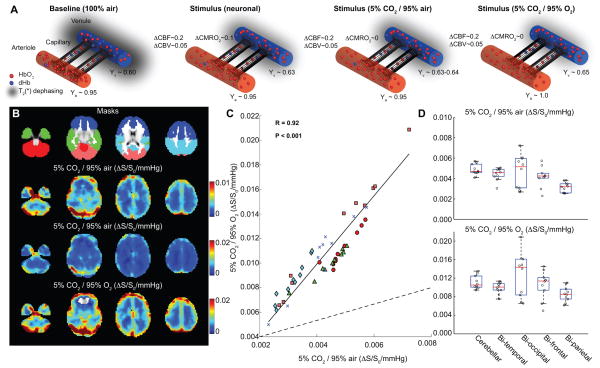
Background and purpose: A promising method for identifying hemodynamic impairment that may serve as a biomarker for stroke risk in patients with intracranial stenosis is cerebrovascular reactivity (CVR) mapping using noninvasive MRI. Here, abilities to measure CVR safely in the clinic using hypercarbic hyperoxic (carbogen) gas challenges, which increase oxygen delivery to tissue, are investigated.

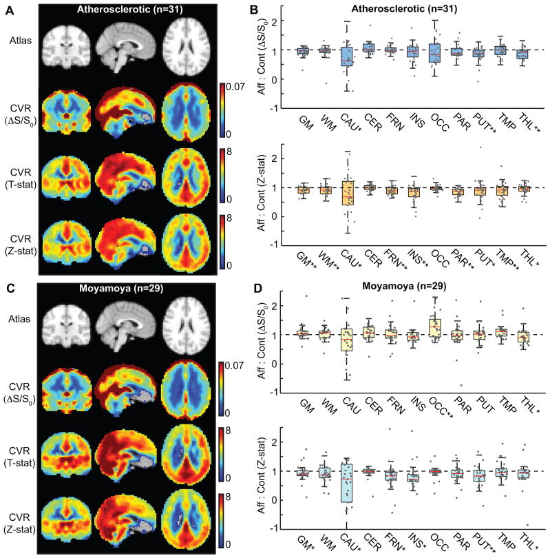
Methods: In sequence with structural and angiographic imaging, blood oxygenation level-dependent carbogen-induced CVR scans were performed in patients with symptomatic intracranial stenosis (n=92) and control (n=10) volunteers, with a subgroup of patients (n=57) undergoing cerebral blood flow-weighted pseudocontinuous arterial spin labeling CVR. Subjects were stratified for 4 substudies to evaluate relationships between (1) carbogen and hypercarbic normoxic CVR in healthy tissue (n=10), (2) carbogen cerebral blood flow CVR and blood oxygenation level-dependent CVR in intracranial stenosis patients (n=57), (3) carbogen CVR and clinical measures of disease in patients with asymmetrical intracranial atherosclerotic (n=31) and moyamoya (n=29) disease, and (4) the CVR scan and immediate and longer-term complications (n=92).

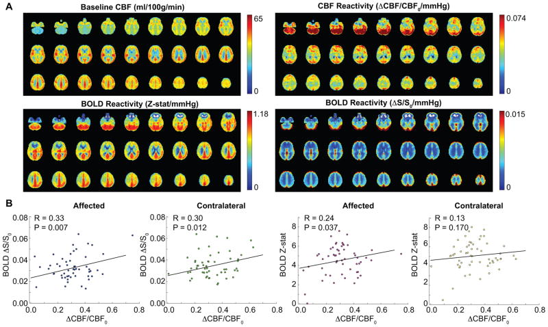
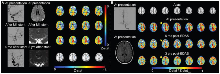
Results: Noninvasive blood oxygenation level-dependent carbogen-induced CVR values correlate with (1) lobar hypercarbic normoxic gas stimuli in healthy tissue (R=0.92; P<0.001), (2) carbogen-induced cerebral blood flow CVR in patients with intracranial stenosis (R=0.30-0.33; P<0.012), and (3) angiographic measures of disease severity both in atherosclerotic and moyamoya patients after appropriate processing. No immediate stroke-related complications were reported in response to carbogen administration; longer-term neurological events fell within the range for expected events in this patient population.

Conclusions: Carbogen-induced CVR elicited no added adverse events and provided a surrogate marker of cerebrovascular reserve consistent with intracranial vasculopathy.

Keywords: constriction, pathologic; hypercapnia; magnetic resonance imaging; regional blood flow; stroke.


Figures

Figure 1
Figure 1. Hypercarbic gas comparison
(A) Graphical representations of fractional changes in oxy-hemoglobin (HbO2) relative to deoxy-hemoglobin (dHb) at baseline and for different stimuli. For neuronal stimuli, cerebral blood flow (CBF), cerebral blood volume (CBV) and the cerebral metabolic rate of oxygen consumption (CMRO2) all increase, causing a small increase in venous oxygenation (Yv) in veins. For 5%CO2/95%air, CBF and CBV increase only. For hypercarbic hyperoxia (i.e., carbogen; 5%CO2/95%O2), CBF and CBV increase, however increases in the partial pressure of arterial oxygen will lead to elevation of arterial (Ya) and Yv, increasing the BOLD effect in a manner non-specific to cerebrovascular reactivity (CVR). (B) Signal changes (ΔS/S0) normalized by change in end-tidal CO2 for the different gas stimuli for healthy volunteers; the hypercarbic normoxic maps have been scaled for maximal contrast and identically to carbogen stimuli maps (Table 1; n=10). (C) The hypercarbic normoxic Δ S/S0/mmHg is smaller than the carbogen ΔS/S0/mmHg as expected, however the two measures correlate tightly (P<0.05) across all brain lobes; colors denote different lobes shown in (B; Masks). Dashed line=line of unity; solid line=best fit line to data. The boxplot in (D) also demonstrate that relative ΔS/S0 between brain regions vary similarly for the two gas stimuli. These data are consistent with carbogen BOLD CVR providing similar information to hypercarbic normoxic BOLD CVR, however with an additional contribution from increased oxygen saturation secondary to hyperoxia.
Figure 2
Figure 2. (ii) Cerebral blood flow (CBF) vs. blood oxygenation level-dependent (BOLD) cerebrovascular reactivity (CVR)
(A) Baseline and CBF CVR (upper) and BOLD CVR (Z-stat/mmHg and ΔS/S0/mmHg) for the same patients (Table 1; n=57). (B) The relationship between BOLD and CBF CVR for all gray matter in affected and contralateral hemispheres. Correlations are found between both measures of CVR for all scenarios except for BOLD Z-stat vs. CBF CVR in contralateral hemisphere.
Figure 3
Figure 3. Blood oxygenation level-dependent (BOLD) cerebrovascular reactivity (CVR) and lateralizing disease
CVR maps calculated according to signal changes (ΔS/S0), T-statistic (T-stat), and Z-statistic (Z-stat). Maps for affected:contralateral ratios for (A,B) atherosclerotic (n=31) and (C,D) moyamoya (n=29) patients. Data were quantified in different regions based on anatomical atlases: GM=total gray matter; WM=total white matter; CAU=caudate; CER=cerebellum; FRN=frontal gray matter; INS=insula; OCC=occipital gray matter; PAR=parietal gray matter; PUT=putamen; TMP=temporal gray matter; THL=thalamus. *P<0.05; **P<0.0045 (Bonferroni-corrected P-value; comparisons=11).
Figure 4
Figure 4. Example patient images
(A) Atherosclerotic patient example. Digital subtraction angiography (DSA) demonstrates severe left M1 stenosis, which is successfully treated with stent placement. Four months after stent placement, in-stent restenosis is significant. Diffusion weighted imaging (DWI) demonstrates watershed infarcts related to severe M1 stenosis. After stent placement, lumenal stenosis is not directly evaluated by computed tomography angiography, but is inferred from decreased opacification of left M2 branches. Blood oxygenation level-dependent (BOLD) cerebrovascular reactivity (CVR) was performed two years after stent placement, when angioplasty was considered due to patient’s increasing dysarthria. CVR is markedly reduced in the left MCA territory with negative z-statistic. (B) Moyamoya patient example. Pre-operatively, although DSA from left internal carotid artery (ICA) injection demonstrates bilateral moyamoya disease (mSS=3 on right; mSS=2 on left), and only prior infarct is in left caudate head on FLAIR, BOLD CVR is markedly reduced in the right cerebral hemisphere, thus indirect revascularization with encephaloduroarteriosynangiosis (EDAS) was performed on the right. Revascularization response is demonstrated on CVR images over a three-year duration. Responses have been normalized by occipital Z-statistics (Z-statocc) to allow for comparison between time points.

References

    1. Holmstedt CA, Turan TN, Chimowitz MI. Atherosclerotic intracranial arterial stenosis: risk factors, diagnosis, and treatment. Lancet Neurol. 2013;12:1106–1114. - PMC - PubMed
    1. Derdeyn CP, Chimowitz MI, Lynn MJ, Fiorella D, Turan TN, Janis LS, et al. Aggressive medical treatment with or without stenting in high-risk patients with intracranial artery stenosis (SAMMPRIS): the final results of a randomised trial. Lancet. 2014;383:333–341. - PMC - PubMed
    1. Zaidat OO, Castonguay AC, Fitzsimmons BF, Woodward BK, Wang Z, Killer-Oberpfalzer M, et al. Design of the Vitesse Intracranial Stent Study for Ischemic Therapy (VISSIT) trial in symptomatic intracranial stenosis. J Stroke Cerebrovasc Dis. 2013;22:1131–1139. - PubMed
    1. Mazighi M, Tanasescu R, Ducrocq X, Vicaut E, Bracard S, Houdart E, et al. Prospective study of symptomatic atherothrombotic intracranial stenoses: the GESICA study. Neurology. 2006;66:1187–1191. - PubMed
    1. Liebeskind DS, Cotsonis GA, Saver JL, Lynn MJ, Turan TN, Cloft HJ, et al. Collaterals dramatically alter stroke risk in intracranial atherosclerosis. Ann Neurol. 2010;69:963–974. - PMC - PubMed

Publication types

MeSH terms