

Dopaminergic modulation of resting-state functional connectivity in de novo patients with Parkinson's disease
- PMID: 24938993
- PMCID: PMC6869560
- DOI: 10.1002/hbm.22561
Dopaminergic modulation of resting-state functional connectivity in de novo patients with Parkinson's disease
Abstract
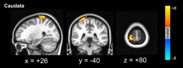
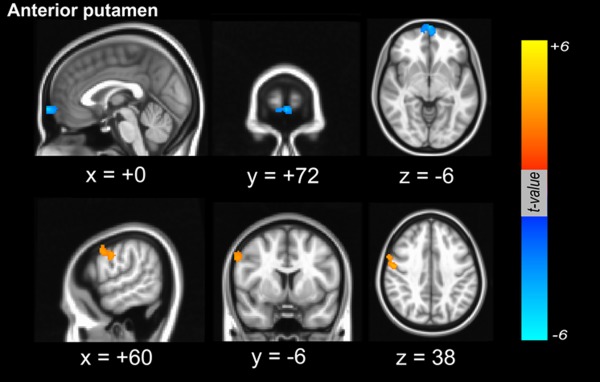
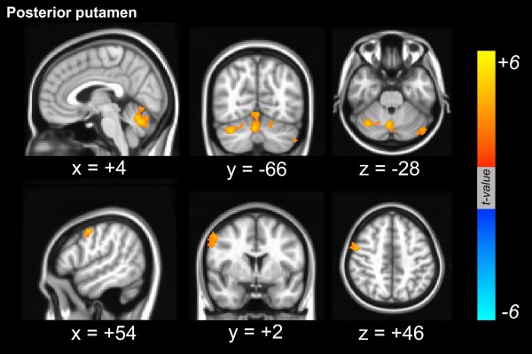
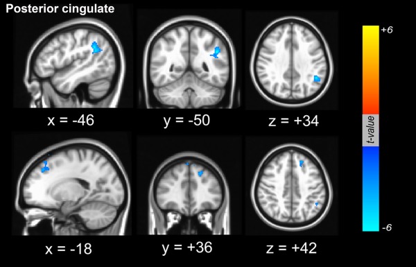
Parkinson's disease (PD) is characterized by degenerative changes of nigral dopamine neurons, resulting in the dopaminergic denervation of the striatum. Resting state networks studies have demonstrated that dopamine modulates distinct network connectivity patterns in both a linear and a nonlinear fashion, but quantitative analyses of dopamine-dependent functional connectivity secondary to PD pathology were less informative. In the present study, we performed a correlation analysis between striatal dopamine levels assessed quantitatively by FP-CIT positron emission tomography imaging and resting-state functional connectivity in 23 drug naïve de novo patients with PD to elucidate dopamine-dependent functional networks. The major finding is that the patterns of dopamine-dependent positive functional connectivity varied depending on the location of striatal seeds. Dopamine-dependent functional connectivity with the caudate predominantly overlay pericentral cortical areas, whereas dopamine-dependent structures functionally connected with the posterior putamen predominantly involved cerebellar areas. The dorsolateral frontal area overlapped as a dopamine-dependent cortical region that was positively connected with the anterior and posterior putamen. On the other hand, cortical areas where functional connectivity from the posterior cingulate was negatively correlated with dopaminergic status in the posterior putamen were localized in the left anterior prefrontal area and the parietal area. Additionally, functional connectivity between the anterior putamen and mesiofrontal areas was negatively coupled with striatal dopamine levels. The present study demonstrated that dopamine-dependent functional network connectivity secondary to PD pathology mainly exhibits a consistent pattern, albeit with some variation. These patterns may reflect the diverse effects of dopaminergic medication on parkinsonian-related motor and cognitive performance.
Keywords: de novo Parkinson's disease; dopamine; resting-state functional connectivity.
Copyright © 2014 Wiley Periodicals, Inc.
Figures
References
-
- Aarsland D, Andersen K, Larsen JP, Lolk A, Kragh‐Sorensen P (2003): Prevalence and characteristics of dementia in Parkinson disease: An 8‐year prospective study. Arch Nurol 60:387–392. - PubMed
-
- Aarsland D, Bronnick K, Williams‐Gray C, Weintraub D, Marder K, Kulisevsky J, Burn D, Barone P, Pagonabarraga J, Allcock L, Santangelo G, Foltynie T, Janvin C, Larsen JP, Barker RA, Emre M (2010): Mild cognitive impairment in Parkinson disease: A multicenter pooled analysis. Neurology 75:1062–1069. - PMC - PubMed
Publication types
MeSH terms
Substances
LinkOut - more resources
Full Text Sources
Other Literature Sources
