

Fractures of the ankle joint: investigation and treatment options
- PMID: 24939377
- PMCID: PMC4075279
- DOI: 10.3238/arztebl.2014.0377
Fractures of the ankle joint: investigation and treatment options
Abstract
Background: Ankle fractures are common, with an incidence of up to 174 cases per 100 000 adults per year. Their correct classification and treatment are of decisive importance for clinical outcome.
Method: Selective review of the literature.
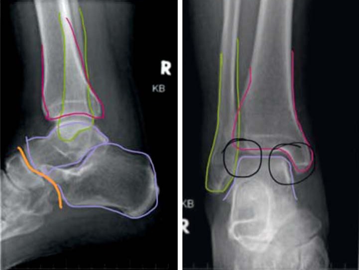
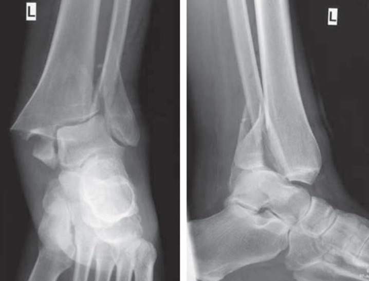
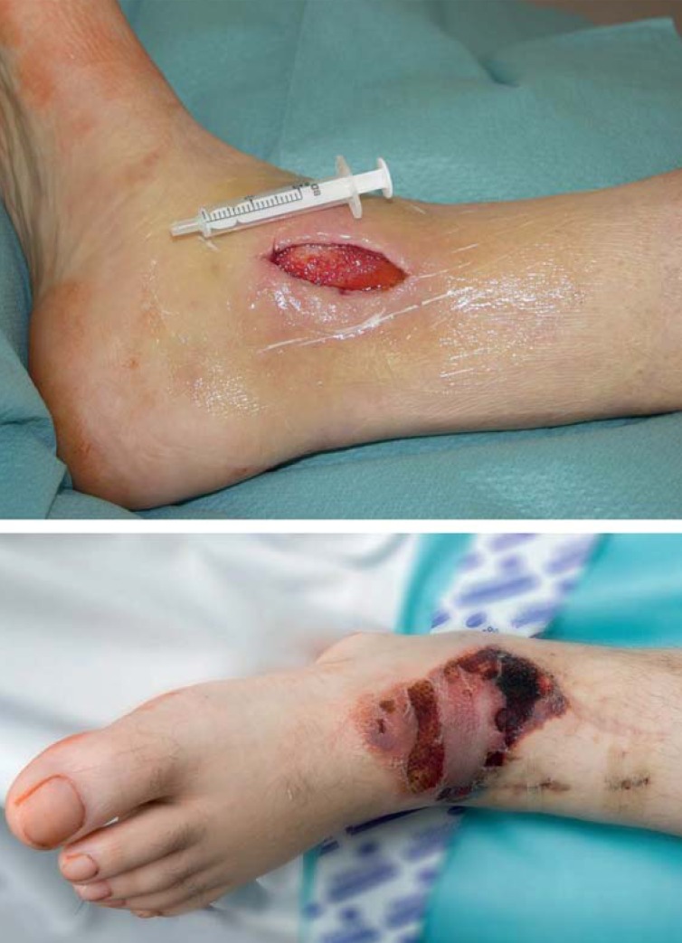
Results: Ankle fractures are initially evaluated by physical examination and then by x-ray. They can be classified according to either the AO Foundation (Association for the Study of Internal Fixation) or the Weber classification. Dislocated fractures need emergency treatment with immediate reduction; this is crucial for the prevention of hypoperfusion and nerve damage. Weber A fractures can usually be treated conservatively, while Weber B and C fractures are usually treated with surgery. An evaluation of the stability of the syndesmosis is important for anatomical reconstruction of the joint. Wound hematoma and wound-edge necrosis are the most common complications, and the postoperative infection rate is 2%. Up to 10% of patients develop ankle arthrosis over the intermediate or long term.
Conclusion: With properly chosen treatment, a good clinical outcome can be achieved. The long-term objective is to prevent post-traumatic ankle arthrosis. The evidence level for optimal treatment strategies is low.
Figures
Comment in
-
Put the "walker" on.Dtsch Arztebl Int. 2014 Oct 17;111(42):721. doi: 10.3238/arztebl.2014.0721a. Dtsch Arztebl Int. 2014. PMID: 25385484 Free PMC article. No abstract available.
-
Contradictory weight-bearing recommendations.Dtsch Arztebl Int. 2014 Oct 17;111(42):721-2. doi: 10.3238/arztebl.2014.0721b. Dtsch Arztebl Int. 2014. PMID: 25385485 Free PMC article. No abstract available.
-
In reply.Dtsch Arztebl Int. 2014 Oct 17;111(42):722. doi: 10.3238/arztebl.2014.0722a. Dtsch Arztebl Int. 2014. PMID: 25385486 Free PMC article. No abstract available.
References
-
- Kannus P, Palvanen M, Niemi S, Parkkari J, Jarvinen M. Increasing number and incidence of low-trauma ankle fractures in elderly people: Finnish statistics during 1970-2000 and projections for the future. Bone. 2002;31:430–433. - PubMed
-
- Pretterklieber ML. Anatomie und Kinematik der Sprunggelenke des Menschen. Der Radiologe. 1999;39:1–7. - PubMed
-
- Thordarson DB, Motamed S, Hedman T, Ebramzadeh E, Bakshian IS. The effect of fibular malreduction on contact pressures in an ankle fracture malunion model. J Bone Joint Surg Am. 1997;79:1809–1815. - PubMed
-
- Ramsey PL, Hamilton W. Changes in tibiotalar area of contact caused by lateral talar shift. J Bone Joint Surg Am. 1976;58:356–357. - PubMed
Publication types
MeSH terms
LinkOut - more resources
Full Text Sources
Other Literature Sources
Medical