. 2014 Oct;69(10):1300-7.
doi: 10.1111/all.12446. Epub 2014 Jun 17.

Group 2 innate lymphoid cells and CD4+ T cells cooperate to mediate type 2 immune response in mice

Affiliations

Group 2 innate lymphoid cells and CD4+ T cells cooperate to mediate type 2 immune response in mice

L Y Drake et al. Allergy. 2014 Oct.

Abstract

Background: Innate lymphoid cells (ILCs) play important roles in innate immunity and tissue remodeling via production of various cytokines and growth factors. Group 2 ILCs (ILC2s) were recently shown to mediate the immune pathology of asthma even without adaptive immunity. However, little is known about possible interactions between ILC2s and other immune cells. We sought to investigate the capacity of ILC2s to regulate effector functions of T cells.

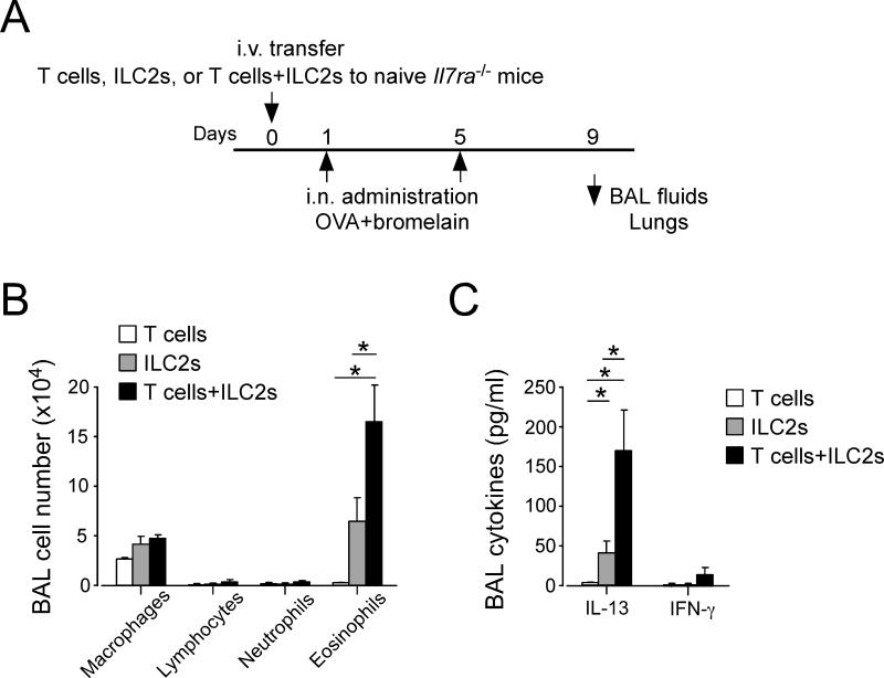
Methods: We isolated ILC2s from the lungs of naïve mice. We cultured CD4(+) T cells with ILC2s in vitro and examined the functions of these cell types. The mechanisms were investigated using blocking antibodies and cells isolated from cytokine-deficient mice. For the in vivo study, we adoptively transferred ILC2s and CD4(+) T cells into Il7ra(-/-) mice and subsequently exposed the mice to ovalbumin and a cysteine protease.

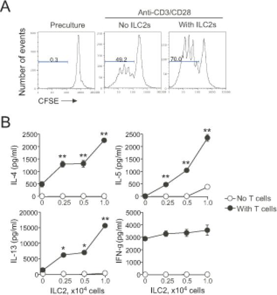
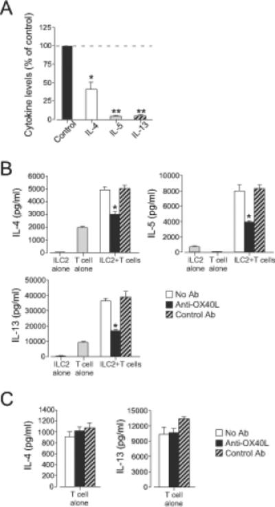
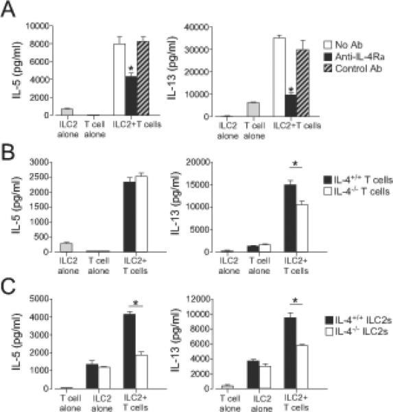
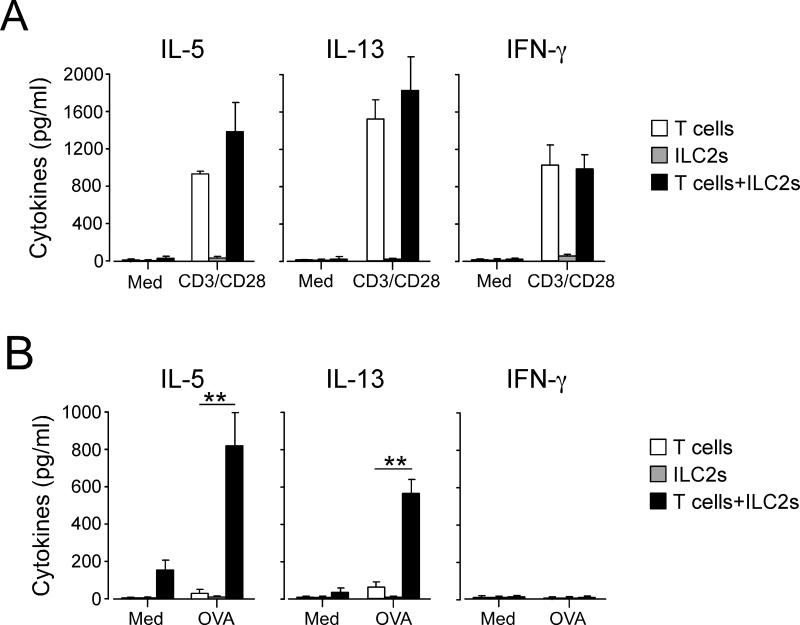
Results: Lung ILC2s enhanced CD4(+) T-cell proliferation and promoted production of type 2 cytokines in vitro. The interaction between ILC2s and CD4(+) T cells involved costimulatory molecule OX40L and cytokine IL-4, which was mainly derived from ILC2s. Adoptive transfer of both ILC2 and CD4(+) T-cell populations, but not each population alone, into Il7ra(-/-) mice resulted in induction of a robust antigen-specific type 2 cytokine response and airway inflammation.

Conclusion: Lung ILC2s function to promote adaptive immunity in addition to their established roles in innate immunity. This novel function of ILC2s needs to be taken into account when considering the pathophysiology of asthma and other allergic airway diseases.

Keywords: T cells; animal models; innate immunity; lymphocytes.


Figures

Figure 1
Figure 1. Co-culture of ILC2s and CD4+ T cells enhances proliferation of CD4+ T cells and type 2 cytokine production
(A) CFSE-labeled CD4+ T cells (2×10⁴ cells/well) were stimulated with anti-CD3/CD28 with or without isolated lung ILC2s (104 cells/well) for 4 days. Cells were analyzed by FACS by gating on CD4+ T cells. Data are representative of three experiments. (B) CD4+ T cells (2×10⁴ cells/well) were stimulated with anti-CD3/CD28 with the indicated numbers of ILC2s for 4 days. Cytokine levels in the supernatants were analyzed by ELISA. *, p<0.05; **, p<0.01 as compared to no ILC2s in the culture. Data (mean±SEM, n=3) are representative of 2-3 experiments.
Figure 2
Figure 2. The interaction between ILC2s and CD4+ T cells is likely bidirectional
(A) CD4+ T cells (5×10⁵ cells/well) and ILC2s (5×10⁵ cells/well) were cultured alone or together for 20 h and then sorted by FACS. Cytokine mRNA expression in each cell population was analyzed. Data are presented as the ratio of mRNA copy numbers in co-cultured cells divided by mRNA copy numbers in cells cultured alone. Data are from three experiments. (B) CD4+ T cells derived from WT or Il5−/− mice were stimulated with anti-CD3/CD28 with or without ILC2s from WT mice for 4 days. *, p<0.05; **, p<0.01 between the groups indicated by horizontal lines. Data (mean±SEM, n=3) are representative of two experiments.
Figure 3
Figure 3. Cellular contact through OX40L plays a key role in the ILC2 and CD4+ T cell interaction
(A) ILC2s (105 cells/well) and CD4+ T cells (2×10⁵ cells/well) were cultured in the Transwell® system for 4 days. Cytokine levels in the supernatants were analyzed by ELISA. Data were normalized to the values from co-culture without the Transwell® system as 100% (control). *, p<0.05; **, p<0.01 as compared to the control. Data (mean±SEM) are a pool of three experiments. (B) ILC2s (104 cells/well) and CD4+ T cells (2×10⁴ cells/well) were stimulated with anti-CD3/CD28 with or without anti-OX40L (10 μg/ml) for 4 days. *, p<0.05 versus no antibody. (C) CD4+ T cells (2×10⁴ cells/well) were stimulated with anti-CD3/CD28 with or without anti-OX40L (10 μg/ml) for 4 days. Data (mean±SEM, n=3) are representative of five experiments.
Figure 4
Figure 4. ILC2-derived IL-4 plays a key role in the ILC2 and CD4+ T cell interaction
(A) ILC2s (104 cells/well) and CD4+ T cells (2x10⁴ cells/well) were stimulated with anti-CD3/CD28 with or without anti-IL-4Rα(10 μg/ml) for 4 days. Cytokine levels in the supernatants were analyzed by ELISA. *, p<0.05 versus no antibody. (B) ILC2s from WT mice and CD4+ T cells from WT or Il4−/− mice were cultured alone or together for 4 days. *, p<0.05 between the groups indicated by horizontal bars. (C) CD4+ T cells from WT mice and ILC2s from WT or Il4−/− mice were cultured alone or together for 4 days. *, p<0.05 between the groups indicated by horizontal bars. Data (mean±SEM, n=3) are representative of three experiments.
Figure 5
Figure 5. ILC2s and CD4+ T cells synergize in antigen-induced airway inflammation in vivo
(A) Experimental protocol. Naïve Il7ra−/− mice were adoptively transferred with CD4+ T cells, ILC2s or both. Mice were then exposed i.n. to OVA plus an adjuvant protease (bromelain) on days 1 and 5 and analyzed on day 9. (B) Cell numbers and differentials in BAL fluids were analyzed and presented as mean±SEM (n =3-4). *, p<0.05 between the groups indicated by horizontal lines. Data are representative of two experiments. (C) Cytokine levels in BAL fluids were analyzed by ELISA and presented as mean±SEM (n =3-4). *, p<0.05 between the groups indicated by horizontal lines. Data are representative of two experiments.
Figure 6
Figure 6. Antigen-specific immune response in CD4+ T cells is enhanced when ILC2s are transferred together with CD4+ T cells
(A) Naïve Il7ra−/− mice were treated as described in Fig. 5A. Lung cells were collected on day 9 and cultured with medium alone or anti-CD3/CD28 antibodies for 5 days. Cytokine levels in the supernatants were analyzed by ELISA. (B) Lung cells were cultured with medium alone or OVA antigen (100 μg/ml) for 7 days. Cytokine levels in the supernatants were analyzed by ELISA. **, p<0.01 between the groups indicated by horizontal lines. Data are representative of two experiments.

Comment in

References

    1. Spits H, Cupedo T. Innate lymphoid cells: emerging insights in development, lineage relationships, and function. Annu. Rev. Immunol. 2012;30:647–675. - PubMed
    1. Walker JA, McKenzie AN. Development and function of group 2 innate lymphoid cells. Curr. Opin. Immunol. 2013;25:148–55. - PMC - PubMed
    1. Neill DR, Wong SH, Bellosi A, Flynn RJ, Daly M, Langford TK, et al. Nuocytes represent a new innate effector leukocyte that mediates type-2 immunity. Nature. 2010;464:1367–1370. - PMC - PubMed
    1. Saenz SA, Siracusa MC, Perrigoue JG, Spencer SP, Urban JF, Jr, Tocker JE, et al. IL-25 elicits a multipotent progenitor cell population that promotes Th2 cytokine responses. Nature. 2010;464:1362–1366. - PMC - PubMed
    1. Saenz SA, Siracusa MC, Monticelli LA, Ziegler CG, Kim BS, Brestoff JR, et al. IL-25 simultaneously elicits distinct populations of innate lymphoid cells and multipotent progenitor type 2 (MPPtype2) cells. J. Exp. Med. 2013;210:1823–1837. - PMC - PubMed

Publication types