. 2014 Jun 15;5(11):3526-40.
doi: 10.18632/oncotarget.1954.

Acrolein- and 4-Aminobiphenyl-DNA adducts in human bladder mucosa and tumor tissue and their mutagenicity in human urothelial cells

Affiliations

Acrolein- and 4-Aminobiphenyl-DNA adducts in human bladder mucosa and tumor tissue and their mutagenicity in human urothelial cells

Hyun-Wook Lee et al. Oncotarget. .

Abstract

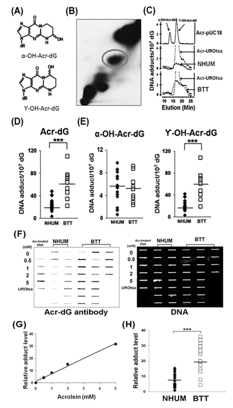
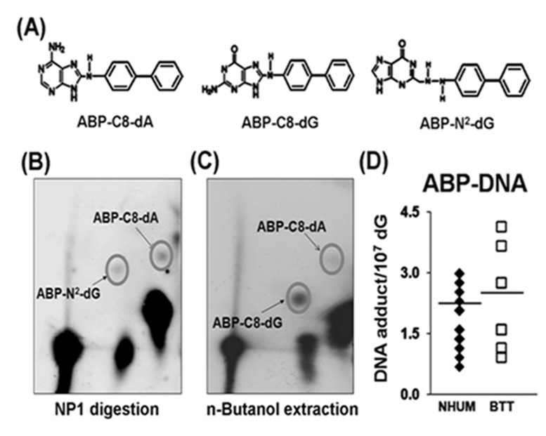
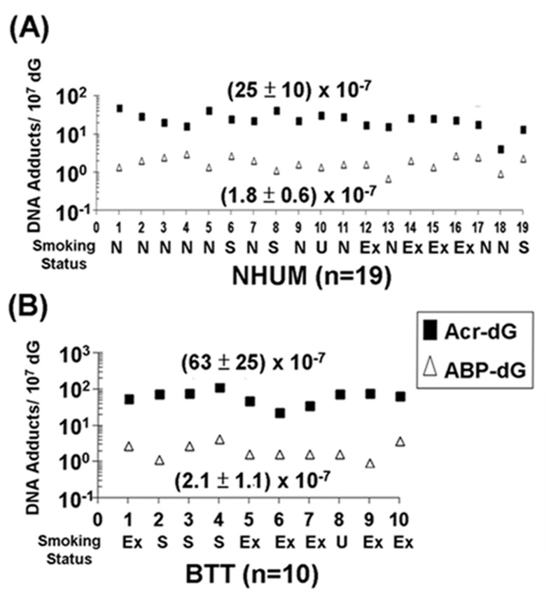
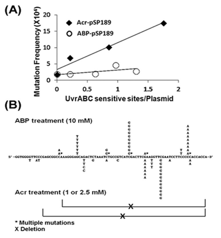
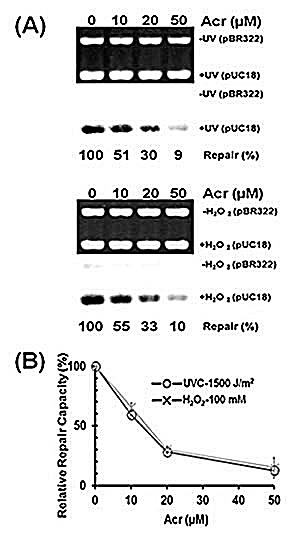
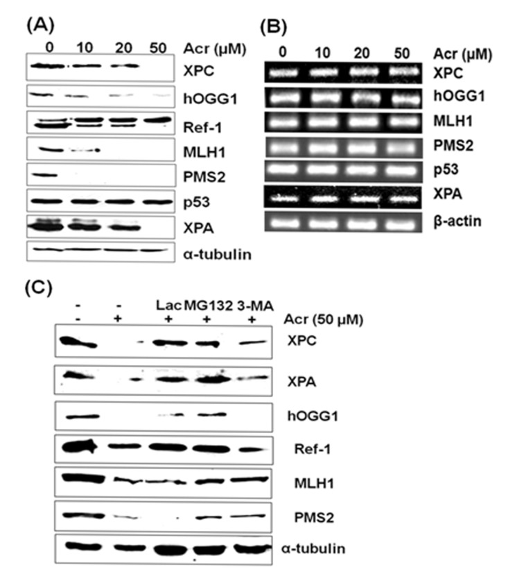
Tobacco smoke (TS) is a major cause of human bladder cancer (BC). Two components in TS, 4-aminobiphenyl (4-ABP) and acrolein, which also are environmental contaminants, can cause bladder tumor in rat models. Their role in TS related BC has not been forthcoming. To establish the relationship between acrolein and 4-ABP exposure and BC, we analyzed acrolein-deoxyguanosine (dG) and 4-ABP-DNA adducts in normal human urothelial mucosa (NHUM) and bladder tumor tissues (BTT), and measured their mutagenicity in human urothelial cells. We found that the acrolein-dG levels in NHUM and BTT are 10-30 fold higher than 4-ABP-DNA adduct levels and that the acrolein-dG levels in BTT are 2 fold higher than in NHUM. Both acrolein-dG and 4-ABP-DNA adducts are mutagenic; however, the former are 5 fold more mutagenic than the latter. These two types of DNA adducts induce different mutational signatures and spectra. We found that acrolein inhibits nucleotide excision and base excision repair and induces repair protein degradation in urothelial cells. Since acrolein is abundant in TS, inhaled acrolein is excreted into urine and accumulates in the bladder and because acrolein inhibits DNA repair and acrolein-dG DNA adducts are mutagenic, we propose that acrolein is a major bladder carcinogen in TS.


Figures

Figure 1
Figure 1. Acrolein (Acr)-dG DNA adduct analysis in normal human urothelial mucosa (NHUM) and bladder tumor tissue (BTT) samples
(A) Chemical structures of α-OH-acrolein-dG and γ-OH-acrolein-dG. Genomic DNA from normal human urothelial mucosa and bladder tumor tissues were prepared and the acrolein-dG DNA adduct levels were determined by both a 32P-postlabeling two dimensional TLC/HPLC method (B to E) and by an immunochemical method (F to H) as described (13, 14). (B) A typical two dimensional TLC separation profile of isomeric acrolein-dG DNA adducts formed in normal human urothelial mucosa. The acrolein-dG DNA adducts spots (circled) resolved by two dimensional TLC (B) were extracted and further separated by HPLC (C). Similar results were observed for bladder tumor tissues. (C) The HPLC profiles of DNA adducts from acrolein-modified plasmid pUC18, acrolein-treated UROtsa cells, normal human urothelial mucosa, and bladder tumor tissues were compared. (D & E) Levels of total acrolein-dG (α-OH-acrolein-dG and γ-OH-acrolein-dG adducts) in normal human urothelial mucosa [mean ± s.d. = (25±10) X10−7/dG, n=19] and in bladder tumor tissues [mean ± s.d. = (63 ± 25) × 10−7/dG, n=10. Bars represent the mean value; statistical significance was analyzed with Student T-test. *** represents p value < 0.001. Genomic DNA from normal human urothelial mucosa (NHUM) (NB-2, NB-17 to NB-74, n=16) and bladder tumor tissue (BTT) (BT-1 to BT-75, n=20) used for acrolein-dG DNA adduct analysis by the 32P-postlabeling and two dimensional TLC/HPLC method were used for acrolein-dG DNA adduct detection by an acrolein-dG primary antibody and a quantum dot labeled second antibody in a slot blot apparatus as described [14]. (F) A typical slot blot result is shown. DNA was spotted on the membrane, hybridized with the acrolein-dG antibody, and then the quantum dot conjugated second antibody: first lane, plasmid pUC18 DNA modified with different concentrations of acrolein and DNA isolated from cultured UROtsa cells; second and third lanes, DNA from normal human urothelial mucosa samples; fourth, fifth and sixth lanes, DNA from bladder tumor tissue samples. Left panel, fluorescent development; right panel, the same amounts of DNA loaded in the membrane were stained with methylene blue. (G) Standard calibration curve as determined by fluorescence intensity of relative acrolein-dG DNA adduct level in plasmid pUC18 DNA modified with different concentrations of acrolein. (H) Relative acrolein-dG DNA adduct levels in normal human urothelial mucosa (n=16) and bladder tumor tissue (n=20) samples as detected by the immunochemical method as described above. Note: acrolein-dG DNA adduct levels in 10 BTT samples (BT-1 to BT-33) were detected by both 32P-postlabeling and the immunochemical methods. Because of the low amount of sample, 10 BTT samples (BT-54 to BT-75) were detected by immunochemical method only.
Figure 2
Figure 2. 4-ABP-DNA adduct analysis in normal human urothelial mucosa (NHUM) and bladder tumor tissue (BTT) samples
(A) Chemical structures of three 4-ABP-DNA adducts: 4-ABP-C8-dG, 4-ABP-N2-dG and 4-ABP-C8-dA. The same genomic DNA samples isolated from the normal human urothelial mucosa and bladder tumor tissues that used for acrolein-dG DNA adduct analysis were used for 4-ABP-DNA adduct analysis by the methods described in the text. (B) A typical three dimensional TLC separation of resultant nucleotides from DNA digested with nuclease P1 (NP1). This method is specifically for 4-ABP-N2-dG and 4-ABP-C8-dA adduct analysis. (C) A typical three dimensional TLC result from n-butanol extractions. This method is specifically for 4-ABP-C8-dG adduct analysis. (D) Levels of 4-ABP-DNA adducts in normal human urothelial mucosa [mean ± s.d. = (1.8±0.6) X10−7/dG, n=19] and bladder tumor tissue [mean ± s.d. =(2.1±1.1) × 10−7/dG, n=10], P = 0.32.
Figure 3
Figure 3. Relative levels of acrolein (Acr)-dG DNA adducts and 4-ABP-DNA adducts in normal human urothelial mucosa (NHUM) and bladder tumor tissue (BTT) samples
The methods for acrolein-dG and 4-ABP-DNA adducts are described in Figs. 1 & 2. Two isomeric acrolein-dG DNA adducts and three 4-ABP-DNA adducts were quantified, as previously described [25, 32, 33]. (A) The levels of total acrolein-dG DNA adducts versus the levels of total 4-ABP-DNA adducts in normal human urothelial mucosa (n=19); and (B) the levels of total acrolein-dG DNA adducts versus the levels of total 4-ABP-DNA adducts in bladder tumor tissue (n=10). Each number along the X-axis represents a different individual (NB-2 to NB-74; BT-1 to BT-33). The tobacco smoking status of these individuals is indicated below the number with N represents non-smokers, S represents smokers, U represents unknown, and Ex represents ex-smokers. The amounts of tobacco consumption and smoking history of smokers are unknown. Note: acrolein-dG DNA adduct levels are 10-30 fold higher than 4-ABP-DNA adduct levels in normal human urothelial mucosa and bladder tumor tissues. Acrolein-dG DNA adduct levels are higher (>2 fold) in bladder tumor tissues than in normal human urothelial mucosa (p<0.001). No significant difference was observed in 4-ABP-DNA adduct levels between normal human urothelial mucosa and bladder tumor tissues (p = 0.32).
Figure 4
Figure 4. Relative mutagenicity, mutational signature and mutational spectrum induced by acrolein (Acr)-dG DNA adducts versus 4-ABP-DNA adducts in human urothelial cells
The supF containing plasmid pSP189 DNA was either reacted with acrolein or N-OH-4-ABP and the DNA adduct number per supercoiled plasmid DNA was determined by a UvrABC incision method (Supplementary Figure S3) [25]. Modified plasmids were transfected into UROtsa cells, mutations in the supF gene were detected, quantified, and sequenced as previously described [25, 31]. (A) Mutation frequency per adduct induced by acrolein-dG DNA adducts and 4-ABP-DNA adducts. (B) Mutational signature and spectrum induced by acrolein-dG DNA adducts and 4-ABP-DNA adducts in the supF gene. Note: acrolein-dG DNA adducts are 5-6 fold more mutagenic than 4-ABP-DNA adducts. Both mutational signature (also see Table 1) and spectrum induced by acrolein-dG DNA adducts and 4-ABP-DNA adducts are different.
Figure 5
Figure 5. Acrolein treatment inhibits nucleotide excision repair and base excision repair in human urothelial cells
Immortalized human urothelial (UROtsa) cells were treated with 0, 10, 20, or 50 μM acrolein for 1 h at 37 °C. The nucleotide excision and base excision repair capacity in these cells were determined by (A) an in vitro DNA damage dependent repair synthesis assay using UV-irradiated or H2O2 modified pUC18 plasmid DNA as substrates, and (B) a host cell reactivation assay using UV-irradiated or H2O2 modified luciferase plasmid pGL3 as substrates, as previously described (13, 26).
Figure 6
Figure 6. Effect of acrolein treatment on protein and mRNA levels of XPA, XPC, hOGG1, MLH1, PMS2, and Ref-1 gene products in urothelial cells
UROtsa cells were treated with different concentrations of acrolein for 1 h at 37 °C, the proteins were separated by SDS-PAGE and detected by Western blot (A), and the mRNA levels were detected by RT-PCR (B). In (C) proteosome inhibitor Lactacystin (10 μM) and MG132 (20 μM) and autophagosome inhibitor 3-methyladenine (10 mM) was added to UROtsa cells before acrolein treatment and the proteins were detected. Note: 1) Acrolein treatment induces a dose dependent reduction of XPA, XPC, hOGG1, MLH1 and PMS2 proteins but not p53 and α-tubulin; 2) Proteosome inhibitor Lac and MG132 inhibits the reduction of XPA, XPC, hOGG1, Ref-1, MLH1 and PMS2 proteins induced by acrolein treatments. Autophagosome inhibitor 3-MA partially inhibits the reduction of XPA, XPC, Ref-1, MLH1 and PMS2.

References

    1. Wu XR. Urothelial tumorigenesis: a tale of divergent pathways. Nat Rev Cancer. 2005;5(9):713–725. - PubMed
    1. Siegel R, Naishadham D, Jemal A. Cancer statistics, 2013. CA Cancer J Clin. 2013;63(1):11–30. - PubMed
    1. Freedman ND, Silverman DT, Hollenbeck AR, Schatzkin A, Abnet CC. Association between smoking and risk of bladder cancer among men and women. JAMA. 2011;306(7):737–745. - PMC - PubMed
    1. Talaska G, al-Juburi AZ, Kadlubar FF. Smoking related carcinogen-DNA adducts in biopsy samples of human urinary bladder: identification of N-(deoxyguanosin-8-yl)-4-aminobiphenyl as a major adduct. Proc Natl Acad Sci U S A. 1991;88(12):5350–5354. - PMC - PubMed
    1. Talaska G. Aromatic amines and human urinary bladder cancer: exposure sources and epidemiology. J Environ Sci Health C Environ Carcinog Ecotoxicol Rev. 2003;21(1):29–43. - PubMed

Publication types