. 2014 Jun 17;7(330):ra57.
doi: 10.1126/scisignal.2004838.

A FAK-Cas-Rac-lamellipodin signaling module transduces extracellular matrix stiffness into mechanosensitive cell cycling

Affiliations

A FAK-Cas-Rac-lamellipodin signaling module transduces extracellular matrix stiffness into mechanosensitive cell cycling

Yong Ho Bae et al. Sci Signal. .

Abstract

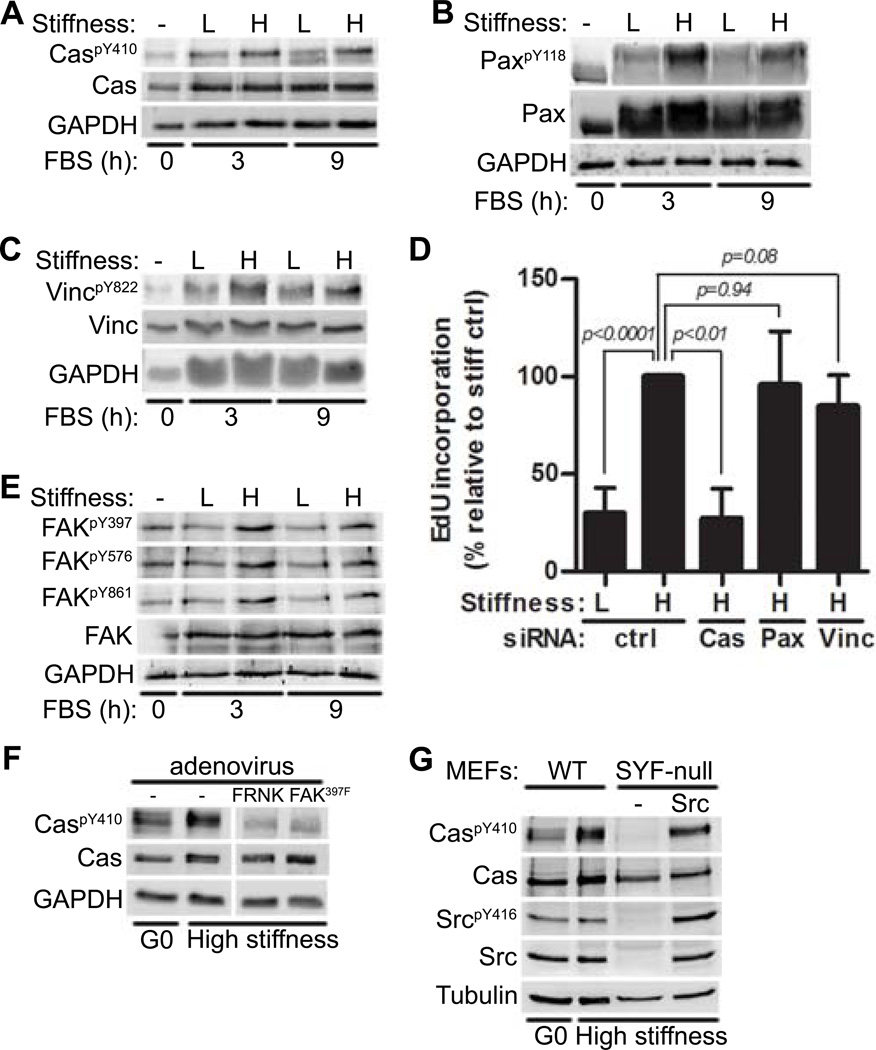
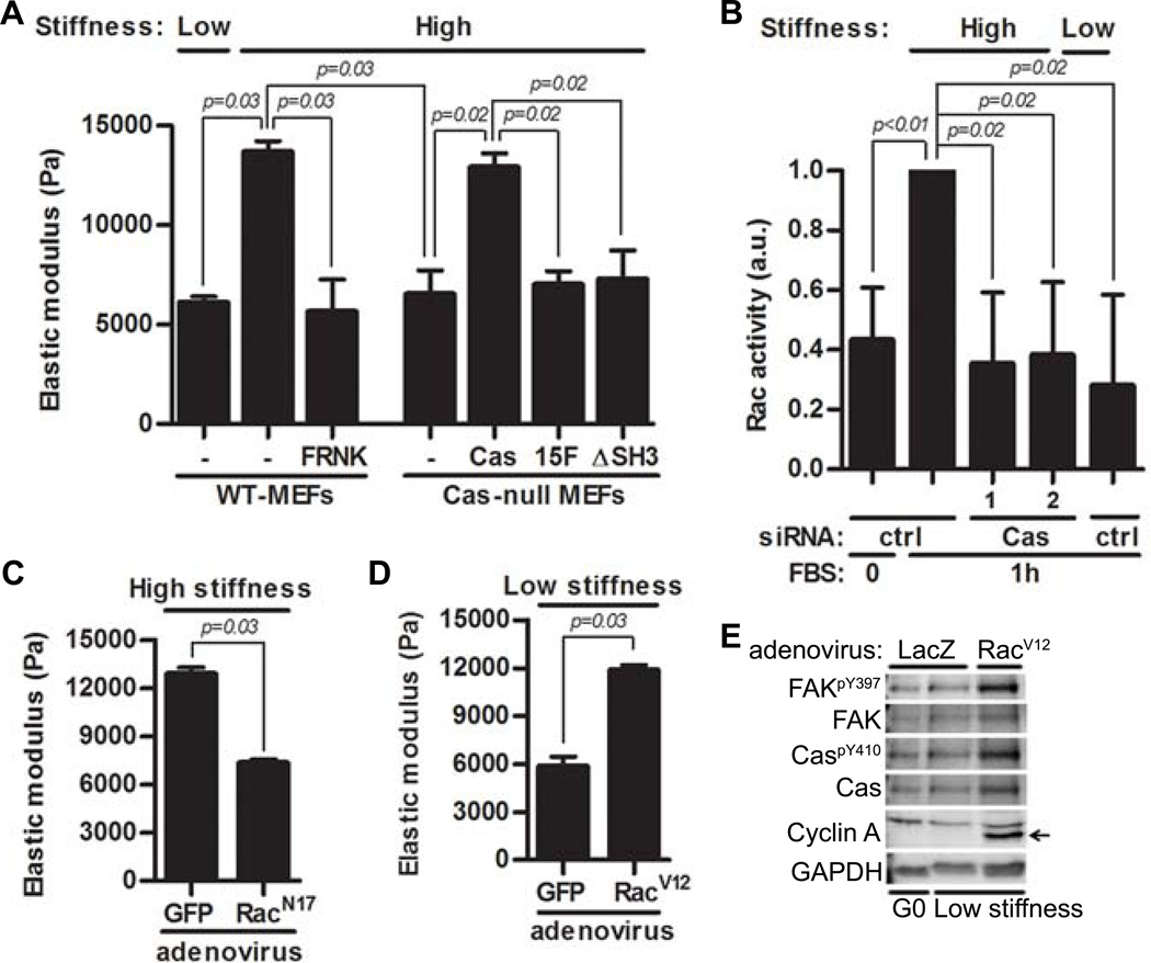
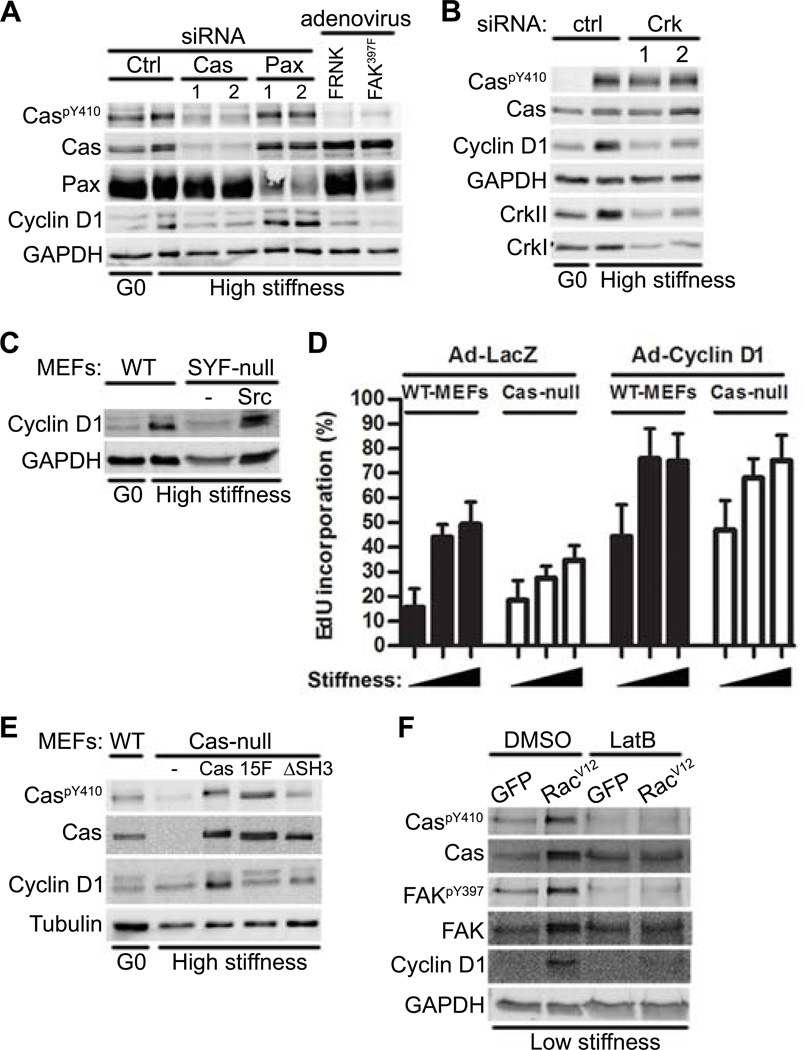
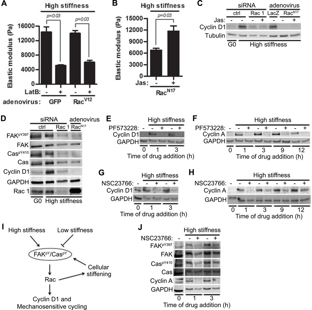
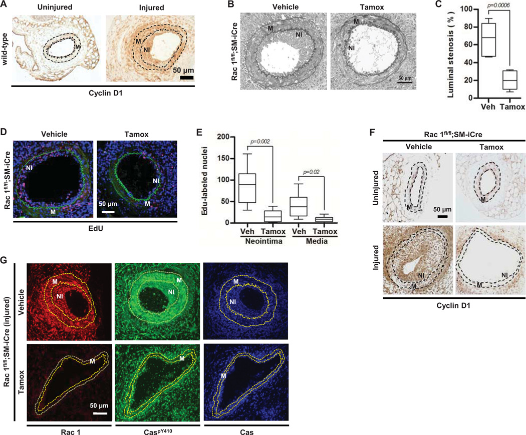
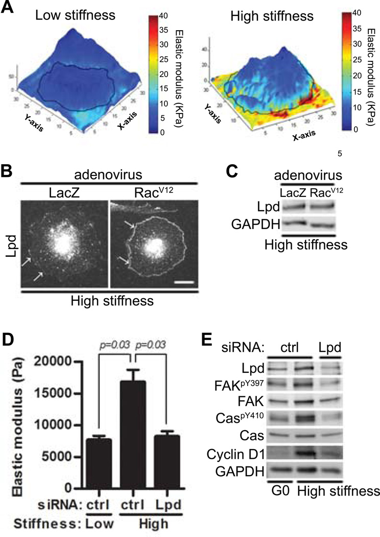
Tissue and extracellular matrix (ECM) stiffness is transduced into intracellular stiffness, signaling, and changes in cellular behavior. Integrins and several of their associated focal adhesion proteins have been implicated in sensing ECM stiffness. We investigated how an initial sensing event is translated into intracellular stiffness and a biologically interpretable signal. We found that a pathway consisting of focal adhesion kinase (FAK), the adaptor protein p130Cas (Cas), and the guanosine triphosphatase Rac selectively transduced ECM stiffness into stable intracellular stiffness, increased the abundance of the cell cycle protein cyclin D1, and promoted S-phase entry. Rac-dependent intracellular stiffening involved its binding partner lamellipodin, a protein that transmits Rac signals to the cytoskeleton during cell migration. Our findings establish that mechanotransduction by a FAK-Cas-Rac-lamellipodin signaling module converts the external information encoded by ECM stiffness into stable intracellular stiffness and mechanosensitive cell cycling. Thus, lamellipodin is important not only in controlling cellular migration but also for regulating the cell cycle in response to mechanical signals.


Figures

Figure 1
Figure 1. FAK/Src-mediated Cas phosphorylation transduces ECM stiffness into mechanosensitive cell cycling
Serum-starved MEFs were incubated on low or high stiffness hydrogels and stimulated with FBS as indicated. Total cell lysates were immunoblotted. (A–C) Lysates analyzed for phosphorylated and total Cas (A), paxillin (B; Pax), or vinculin (C; Vinc). N=3 independent biological replicates for (A) to (C). (D) Starved MEFs transfected with a control (ctrl) siRNA or siRNAs to Cas, paxillin, or vinculin were seeded on high stiffness hydrogels with FBS for 20 hours. S phase entry was assessed by EdU incorporation. Error bars show mean ± SD, N=4 independent biological replicates. (E) Same experiment as in (A) but lysates were probed for Tyr397, Tyr576, Tyr861, and total FAK. (F) Starved MEFs infected with adenoviruses (FRNK or FAK397F) were replated on stiff hydrogels with FBS for 9 hours and probed for phosphorylated and total Cas. The vertical white bar indicates removal of irrelevant lanes from a single blot. N=3 independent biological replicates. (G) WT MEFs, SYF-null (MEFs deficient in Src, Yes, and Fyn) and c-Src-reconstituted SYF-MEFs were plated on high stiffness hydrogels and stimulated with FBS for 9 hours. N=3 independent biological replicates for (E) to (G).
Figure 2
Figure 2. The FAK-Cas-Rac signaling module transduces substratum stiffness into intracellular stiffness and mechanosensitive cell cycling
(A) Serum-starved MEFs and Cas-null MEFs, Cas-null MEFs reconstituted with WT-Cas, Cas15F, CasΔSH3, and FAK-inhibited (FRNK) MEFs were cultured on high or low stiffness hydrogels with FBS. Intracellular stiffness was determined by AFM; error bars show mean ± SD, N=4 independent biological replicates. (B) Starved MEFs transfected with a control (ctrl) siRNA or Cas siRNAs were replated on low- or high stiffness hydrogels with FBS. Rac activity was determined by G-LISA; error bars show mean ± SD, N=4 independent biological replicates. (C) Starved MEFs infected with adenoviruses encoding GFP or RacN17 were plated on high stiffness hydrogels overnight. Intracellular stiffness was determined by AFM; error bars show mean ± SD, N=4 independent biological replicates. (D) Starved MEFs infected with adenoviruses encoding GFP or RacV12 were plated on low stiffness hydrogels with FBS overnight. Intracellular stiffness was determined by AFM; error bars show mean ± SD, N=4 independent biological replicates. (E) Starved MEFs infected with adenoviruses encoding LacZ or RacV12 were plated on low stiffness hydrogels with FBS for 20 hours; lysates were analyzed by immunoblotting. N=3 independent biological replicates.
Figure 3
Figure 3. A FAK-Cas-Rac signaling module is necessary for the mechanosensitive induction of cyclin D1
(A) Immunoblot of starved MEFs transfected with Cas or paxillin siRNAs or infected with adenoviruses encoding FRNK or FAK397F and plated on high or low stiffness hydrogels with FBS for 9 hours. N=3 independent biological replicates. (B) Immunoblot of starved MEFs transfected with control or CrkII siRNAs plated on fibronectin-coated high stiffness hydrogels with FBS for 9 hours. N=3 independent biological replicates. (C) Immunoblot of wild-type MEFs, SYF-null MEFs, and c-Src-reconstituted SYF-MEFs plated on high-stiffness hydrogels with FBS for 9 hours. N=3 independent biological replicates. (D) Immunoblot of wild-type and Cas-null MEFs that were starved, infected with adenoviruses encoding LacZ or Cyclin D1, and plated on low, medium, or high stiffness fibronectin-coated hydrogels. S phase entry was assessed by EdU incorporation; error bars show mean ± SD, N=4 independent biological replicates. (E) Immunoblot of serum-starved wild-type, Cas-null, Cas-reconstituted (Cas), Cas15F, and CasΔSH3 MEFs plated on high stiffness hydrogels with FBS for 9 hours. N=3 independent biological replicates. (F) Immunoblot of starved MEFs infected with adenoviruses encoding GFP or RacV12 and plated on low stiffness hydrogels with FBS ± latrunculin B (LatB) for 9 hours. N=3 independent biological replicates.
Figure 4
Figure 4. Rac-dependent intracellular stiffening maintains information flow through the FAK-Cas-Rac signaling pathway
(A) Starved MEFs infected with adenoviruses encoding GFP or RacV12 were plated on high stiffness hydrogels with FBS ± latrunculin B (LatB); intracellular stiffness was measured by AFM. Error bars show mean ± SD. N=4 independent biological replicates. (B–C) Starved MEFs were either transfected with control or Rac1 siRNA or infected with adenoviruses encoding LacZ or RacN17. The cells were plated on high stiffness hydrogels with FBS ± jasplakinolide (Jas) and analyzed by AFM (B; error bars show mean ± SD) or immunoblotting (C); N=4 and 3 independent biological replicates for (B) and (C), respectively. (D) Immunoblot of starved MEFs infected with adenovirus encoding RacN17 or transfected with control or Rac1 siRNAs. N=3 biological replicates. (E–H) Serum-starved MEFs were plated on high stiffness hydrogels with FBS and treated with the FAK inhibitor PF573228 or the Rac inhibitor NSC23766 at the indicated times. (E and G) Samples were collected after 9 hours, and analyzed by immunoblotting for cyclin D1. N=3 biological replicates. (F and H) The experiment in E and G was repeated except that cells were collected at 20 hours and analyzed for cyclin A. N=3 biological replicates. (I) Model of feed-forward and feed-back mechanotransduction through the FAK-Cas-Rac signaling module. (J) Starved MEFs were seeded on high stiffness hydrogels with FBS and treated with NSC23766 at the indicated times. Lysates were collected at 20 hours (a time when cyclin A would be expressed in cycling cells) and immunoblotted. The vertical white bar indicates removal of irrelevant lanes from a single blot. N=3 independent biological replicates.
Figure 5
Figure 5. Rac1 is essential for the proliferative response to vascular injury in vivo and cyclin D1 abundance and Cas phosphorylation are reduced in the vascular smooth muscle cells of Rac-null arteries after injury
(A) Representative cyclin D1 staining of uninjured and injured femoral artery sections from C57BL/6 mice; N=5. (B) Representative elastin staining of injured femoral artery sections in Rac1flfl;SM-iCre mice treated with vehicle or tamoxifen (tamox). NI; neointima. M; media. (C) Quantification of the percent luminal stenosis; N=7 mice per genotype and treatment. (D) Representative images of EdU incorporation (red) in injured femoral artery sections of Rac1flfl;SM-iCre mice treated with vehicle or tamoxifen. DAPI-stained nuclei and elastic lamina are shown in blue and green respectively. (E) Quantification of the EdU response; N=6–7 mice per genotype and treatment. (F) Cyclin D1 staining of uninjured and injured femoral artery sections from Rac1fl/fl;SM-iCre mice treated with vehicle or tamoxifen. Images are representative of N=3 (uninjured) or N=6 (injured) mice per genotype and treatment. (G) Rac, CaspY410, and total Cas staining of injured femoral artery sections from Rac1fl/fl;SM-iCre mice treated with vehicle or tamoxifen. Images are representative of N=3 mice per genotype and treatment. In (F) and (G), dashed lines show the internal elastic lamina and external elastic laminae. M; media. NI; Neointima. Scale bar = 50 µm.
Figure 6
Figure 6. Lamellipodin links Rac to intracellular stiffening and cell cycling
(A) Starved MEFs were cultured overnight on low or high stiffness hydrogels with FBS. A point-by-point height and stiffness maps of single cells was acquired from AFM-force volume mode measurements. Height images were overlaid with stiffness maps to determine localized mechanical properties for single cells. N=4 independent biological replicates. (B–C) MEFs infected with adenoviruses encoding LacZ or RacV12 plated on high stiffness hydrogels with FBS for 20 hours. (B) Fluorescence images of cells were acquired using Zeiss LSM 510 META/NLO confocal microscope; scale bar= 20 µm. White arrows showed lamellipodin localization at the leading edge. (C) Total cell lysates were analyzed by immunoblotting. N=3 independent biological replicates. (D–E) Starved MEFs transfected with control (ctrl) siRNA or a pool of siRNAs to lamellipodin (Lpd) were plated on low or high stiffness hydrogels with FBS and analyzed by AFM (D) or immunoblotting (E). Error bars show mean ± SD; N=4 (D) and N=3 (E) independent biological replicates.

References

    1. Pelham RJ, Wang Y-l. Cell locomotion and focal adhesions are regulated by substrate flexibility. Proceedings of the National Academy of Sciences. 1997;94:13661–13665. - PMC - PubMed
    1. Paszek M, Weaver V. The Tension Mounts: Mechanics Meets Morphogenesis and Malignancy. Journal of Mammary Gland Biology and Neoplasia. 2004;9:325–342. - PubMed
    1. Engler AJ, Sen S, Sweeney HL, Discher DE. Matrix Elasticity Directs Stem Cell Lineage Specification. Cell. 2006;126:677–689. - PubMed
    1. Provenzano PP, Inman DR, Eliceiri KW, Keely PJ. Matrix density-induced mechanoregulation of breast cell phenotype, signaling and gene expression through a FAK-ERK linkage. Oncogene. 2009;28:4326–4343. - PMC - PubMed
    1. Kothapalli D, Liu S-L, Bae Yong H, Monslow J, Xu T, Hawthorne Elizabeth A, Byfield Fitzroy J, Castagnino P, Rao S, Rader Daniel J, Pure E, Phillips Michael C, Lund-Katz S, Janmey Paul A, Assoian Richard K. Cardiovascular Protection by ApoE and ApoE-HDL Linked to Suppression of ECM Gene Expression and Arterial Stiffening. Cell Reports. 2012;2:1259–1271. - PMC - PubMed

Publication types

Substances

Associated data