. 2014 Jul;42(13):8231-42.
doi: 10.1093/nar/gku525. Epub 2014 Jun 17.

The elusive evidence for chromothripsis

Affiliations

The elusive evidence for chromothripsis

Marcus Kinsella et al. Nucleic Acids Res. 2014 Jul.

Abstract

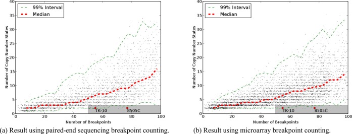
The chromothripsis hypothesis suggests an extraordinary one-step catastrophic genomic event allowing a chromosome to 'shatter into many pieces' and reassemble into a functioning chromosome. Recent efforts have aimed to detect chromothripsis by looking for a genomic signature, characterized by a large number of breakpoints (50-250), but a limited number of oscillating copy number states (2-3) confined to a few chromosomes. The chromothripsis phenomenon has become widely reported in different cancers, but using inconsistent and sometimes relaxed criteria for determining rearrangements occur simultaneously rather than progressively. We revisit the original simulation approach and show that the signature is not clearly exceptional, and can be explained using only progressive rearrangements. For example, 3.9% of progressively simulated chromosomes with 50-55 breakpoints were dominated by two or three copy number states. In addition, by adjusting the parameters of the simulation, the proposed footprint appears more frequently. Lastly, we provide an algorithm to find a sequence of progressive rearrangements that explains all observed breakpoints from a proposed chromothripsis chromosome. Thus, the proposed signature cannot be considered a sufficient proof for this extraordinary hypothesis. Great caution should be exercised when labeling complex rearrangements as chromothripsis from genome hybridization and sequencing experiments.


Figures

Figure 1.
Figure 1.
A hypothetical chromosome of length 100 shattered into blocks of length 10 and then reassembled. The hypothetical rearranged chromosome breakpoints are in Table 1. (a) The breakpoints and block copy number projected onto original chromosome. (b) A breakpoint graph representing the hypothetical rearranged chromosome with Table 1 breakpoints.
Figure 2.
Figure 2.
Following Stephens et al. simulation procedure, a sequence of possible rearrangements steps to explain the observed breakpoints in Table 1.
Figure 3.
Figure 3.
Charts of number of breakpoints versus number of copy number states for simulated chromosomes. The shaded gray area indicates the boundaries of the footprint of chromothripsis proposed by Stephens et al. The cell lines with number of breakpoints and copy number states as described by Stephens et al. are plotted as red points. The red dashed line shows the median number of copy number states for given numbers of breakpoints. The green dashed lines show an interval of copy number states that contains 99% of observations. (a) Results of directly reimplementing the simulation method of Stephens et al. (b) Results after fixing indexing issue for inversions. (c) Results counting breakpoints as they would appear from PES and counting the number of copy number states needed to cover 95% of the chromosome. (d) Results counting breakpoints as they would appear from microarrays or depth of coverage and counting the number of copy number states needed to cover 95% of the chromosome.
Figure 4.
Figure 4.
Charts of breakpoints versus copy number states for simulations with an overrepresentation of inversions. (a) Result using PES breakpoint counting. (b) Result using microarray breakpoint counting.
Figure 5.
Figure 5.
Counts of breakpoints and copy number states from a simulation based on the breakpoints from simulated chromosome in Supplementary Figure S1. The breakpoints and copy number states of the simulated chromosome are indicated on the chart.
Figure 6.
Figure 6.
An illustration of the result of the series of inversions and deletions for chromosome 5 of TK10. The top panel broadly shows the ordering of segments after rearrangement. The upper color bar shows all segments of the unrearranged chromosome colored from blue to red. The lower color bar shows segments with the same coloring after rearrangement. Note that some segments have been deleted so the chromosome is shorter. The middle panel shows the breakpoints achieved by inversions and deletions, and the lower panel shows the observed breakpoints.

Similar articles

Cited by

References

    1. Stephens P.J., Greenman C.D., Fu B., Yang F., Bignell G.R., Mudie L.J., Pleasance E.D., Lau K.W., Beare D., Stebbings L.A., et al. Massive genomic rearrangement acquired in a single catastrophic event during cancer development. Cell. 2011;144:27–40. - PMC - PubMed
    1. Magrangeas F., Avet-Loiseau H., Munshi N.C., Minvielle S. Chromothripsis identifies a rare and aggressive entity among newly diagnosed multiple myeloma patients. Blood. 2011;118:675–678. - PMC - PubMed
    1. Rausch T., Jones D.T., Zapatka M., Stutz A.M., Zichner T., Weischenfeldt J., Jager N., Remke M., Shih D., Northcott P.A., et al. Genome sequencing of pediatric medulloblastoma links catastrophic DNA rearrangements with TP53 mutations. Cell. 2012;148:59–71. - PMC - PubMed
    1. Northcott P.A., Shih D.J., Peacock J., Garzia L., Morrissy A.S., Zichner T., Stutz A.M., Korshunov A., Reimand J., Schumacher S.E., et al. Subgroup-specific structural variation across 1,000 medulloblastoma genomes. Nature. 2012;488:49–56. - PMC - PubMed
    1. Molenaar J.J., Koster J., Zwijnenburg D.A., van Sluis P., Valentijn L.J., van der Ploeg I., Hamdi M., van Nes J., Westerman B.A., van Arkel J., et al. Sequencing of neuroblastoma identifies chromothripsis and defects in neuritogenesis genes. Nature. 2012;483:589–593. - PubMed

Publication types