. 2014 Jun 15;28(12):1291-309.
doi: 10.1101/gad.240291.114.

Tension-dependent removal of pericentromeric shugoshin is an indicator of sister chromosome biorientation

Affiliations

Tension-dependent removal of pericentromeric shugoshin is an indicator of sister chromosome biorientation

Olga O Nerusheva et al. Genes Dev. .

Abstract

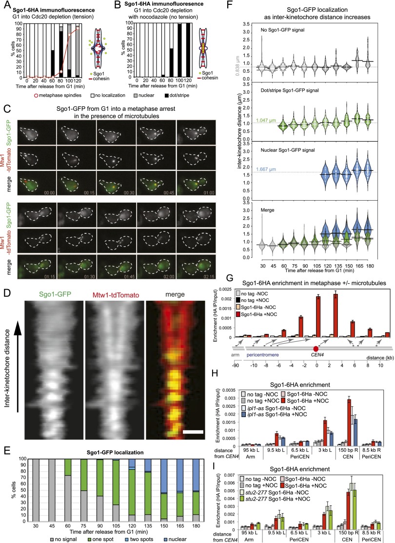
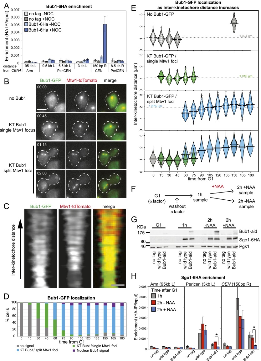
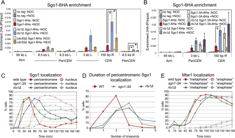
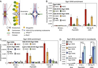
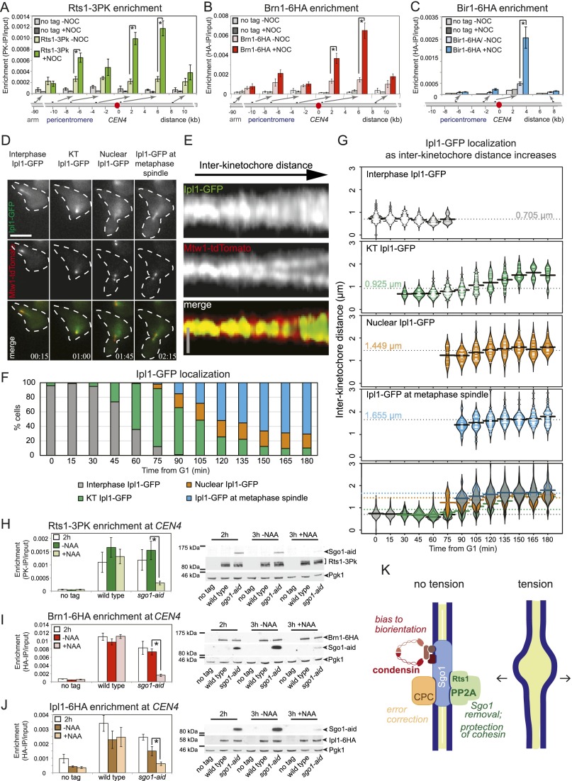
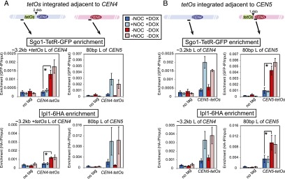
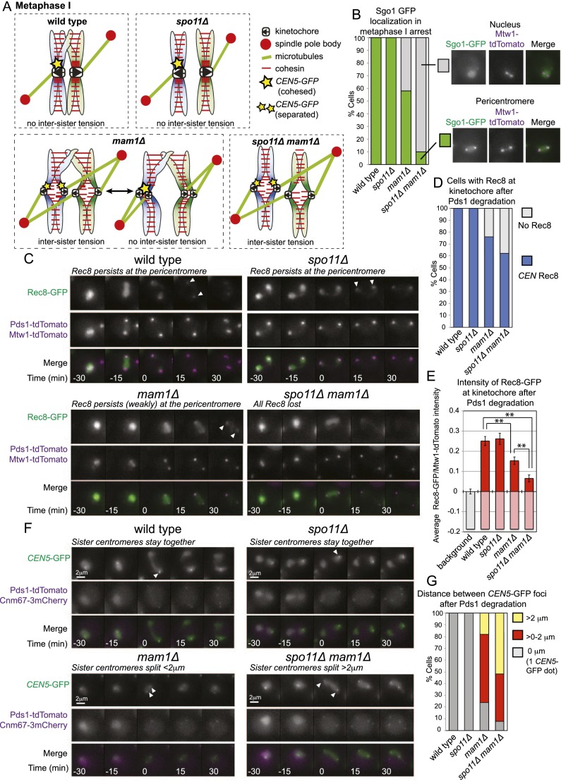
During mitosis and meiosis, sister chromatid cohesion resists the pulling forces of microtubules, enabling the generation of tension at kinetochores upon chromosome biorientation. How tension is read to signal the bioriented state remains unclear. Shugoshins form a pericentromeric platform that integrates multiple functions to ensure proper chromosome biorientation. Here we show that budding yeast shugoshin Sgo1 dissociates from the pericentromere reversibly in response to tension. The antagonistic activities of the kinetochore-associated Bub1 kinase and the Sgo1-bound phosphatase protein phosphatase 2A (PP2A)-Rts1 underlie a tension-dependent circuitry that enables Sgo1 removal upon sister kinetochore biorientation. Sgo1 dissociation from the pericentromere triggers dissociation of condensin and Aurora B from the centromere, thereby stabilizing the bioriented state. Conversely, forcing sister kinetochores to be under tension during meiosis I leads to premature Sgo1 removal and precocious loss of pericentromeric cohesion. Overall, we show that the pivotal role of shugoshin is to build a platform at the pericentromere that attracts activities that respond to the absence of tension between sister kinetochores. Disassembly of this platform in response to intersister kinetochore tension signals the bioriented state. Therefore, tension sensing by shugoshin is a central mechanism by which the bioriented state is read.

Keywords: biorientation; kinetochore; meiosis; mitosis; shugoshin; tension.


Figures

Figure 1.
Figure 1.
Sgo1 is removed from the pericentromere in metaphase in the presence of microtubules. (A,B) Sgo1 dispersal into the nucleus in metaphase is dependent on microtubules. Cells carrying SGO1-6HA and pMET3-CDC20 (strain AM6390) were arrested in G1 with α factor. The culture was split, α factor was washed out, and both cultures were released into medium containing methionine to repress CDC20 and induce arrest in metaphase. Either DMSO (A; tension) or nocodazole (B; no tension) was added. Samples were extracted at the indicated intervals after release from G1 for Sgo1-6HA and tubulin immunofluorescence, and Sgo1 localization (no, dot/stripe, nuclear) and spindle morphology were scored. Schematic diagrams indicate chromosome configuration in the presence (A) or absence (B) of tension. (C) Loss of Sgo1-yeGFP from the pericentromere coincides with the appearance of a bilobed kinetochore signal. Cells carrying SGO1-yeGFP and MTW1-tdTomato (strain AM9233) were imaged on a microfluidics device at 15-min intervals after release from G1 arrest. (D–F) Sgo1-yeGFP loses its pericentromeric localization as kinetochore signals split. Strain AM9233 (pMET3-CDC20 SGO1-yeGFP MTW1-tdTomato) was arrested in G1 using α factor and released in medium containing 8 mM methionine to deplete Cdc20. Images of multiple cells were taken every 15 min, with the first time point taken 0.5 h after the release from G1. (D) Line scans across kinetochore foci of single cells were assembled from 100 images to generate a V plot showing Sgo1-GFP localization as interkinetochore distance increases. Bar, 2μm. (E) Bar chart showing the fraction of cells with the indicated Sgo1 localization at each time point. (F) The distance between Mtw1-tdTomato signals and the localization of Sgo1-yeGFP was scored in 200 cells. The bean plot shows the distribution of interkinetochore distances for which each localization type was scored. The horizontal line represents the mean. (G) Sgo1 is removed from the pericentromere at metaphase in the presence of microtubules. Strains AM6390 (pMET3-CDC20 SGO1-6HA) and AM2508 (pMET3-CDC20; no tag control) were released from G1 into medium containing methionine and either DMSO (−NOC) or nocodazole (+NOC). After 2 h, cells were harvested, and Sgo1-6HA levels at the indicated sites on chromosome IV were analyzed by ChIP-qPCR. The average of three experimental repeats (qPCR performed in triplicate in each case) is shown for AM6390, with error bars representing standard error. For the no tag control (AM2508), representative values are shown from one of these experiments. See also Supplemental Figure S2, G and H, for Sgo1-6HA association with sites on chromosomes III and V. (H) Wild-type (AM6390) and ipl1-as5 (AM8217) cells carrying pMET3-CDC20 and SGO1-6HA as well as a no tag control (AM2508) were treated as in G except that NA-PP1 (50 mM) was added to inhibit Ipl1 when bud formation was observed after release from G1. Sgo1-6HA levels at the indicated sites on chromosome IV were measured by ChIP-qPCR in cells harvested 2 h (wild type) or 2.5 h (ipl1-as) after release from G1 to obtain a similar number of cells arrested in metaphase. (I) The stu2-277 mutation prevents Sgo1 removal in the presence of microtubules. Wild-type (AM6390) and stu2-277 (AM9093) cells carrying pMET3-CDC20 and SGO1-6HA as well as a no tag control (AM2508) were treated as in G except that cells were shifted to 37°C after release from G1. Cells were harvested for Sgo1-6HA ChIP-qPCR after 1.5 h (wild type) or 2.25 h (stu2-277) to obtain similar numbers of cells arrested in metaphase. In H and I, the average of three independent repeats is shown, with error bars representing standard error.
Figure 2.
Figure 2.
Bub1 is removed from kinetochores later than Sgo1 dissociates from the pericentromere. (A) Bub1 associates with centromeres in metaphase-arrested cells only in the absence of spindle tension. Cells (strain AM7449) carrying BUB1-6HA and pMET3-CDC20 and a no tag control (AM2508) were treated as described in Figure 1G. Bub1-6HA levels at the indicated sites were measured by ChIP-qPCR. The average of three experimental repeats is shown, with error bars representing standard error. (B–E) Bub1 is retained at kinetochores upon separation of kinetochore clusters. Cells carrying BUB1-yeGFP and MTW1-tdTomato (strain AM9229) were imaged on a microfluidics device at 15-min intervals after release from G1 arrest. (B) Cells exhibiting different types of Bub1-GFP localization at the indicated time points are shown. Bar, 5 μm. (C) Line scans across kinetochore foci of single cells were assembled from 100 images to generate a V plot showing Bub1-yeGFP localization as interkinetochore distance increases. Bar, 2 μm. (D) Bar chart with the fraction of cells with the indicated Bub1 localization at each time point is shown. (E) The distance between Mtw1-tdTomato signals and the localization of Bub1-yeGFP was scored in at least 90 cells for each time point. The bean plot shows the distribution of interkinetochore distances for which each localization type was scored. Lines within the beans represent individual cells. Beans for small sets of cells (N < 10) are not shown. The horizontal line represents the mean. (F–H) Continued Bub1 presence at kinetochores is required for Sgo1 localization at the pericentromere. (F) Scheme of the experiment is shown. Wild-type (AM6390) and bub1-aid OsTir1 (AM9096) cells carrying SGO1-6HA and a no tag control (AM2508), all carrying pMET3-CDC20, were released from G1 into methionine and nocodazole-containing medium. After 1 h, one-third of the culture was harvested for ChIP and Western blotting, the remaining culture was split, and NAA was added to one half. After 2 h total, the remaining cultures were harvested. (G) Western immunoblot analysis was performed with anti-aid, anti-HA, and anti-Pgk1 antibodies to confirm that Bub1 is degraded upon NAA treatment, but Sgo1 is not. Pgk1 is shown as a loading control. (H) ChIP-qPCR analysis of Sgo1 localization at the indicated sites on chromosome IV. The mean of three experimental repeats is shown, with error bars indicating standard error. Student’s t-test was used to calculate confidence values. (*) P < 0.05.
Figure 3.
Figure 3.
Association with PP2ARts1 is required for timely Sgo1 removal from the pericentromere. (A) Pericentromeric Sgo1 levels are regulated by Rts1 and Cdc55. Wild-type (AM6390), rts1Δ (AM8859), and cdc55Δ (AM8957) cells carrying SGO1-6HA and pMET3-CDC20 and a no tag pMET3-CDC20 control (AM2508) were arrested in metaphase in the presence or absence of microtubules as described in Figure 1G, and anti-HA ChIP was performed followed by qPCR with primer sets at the indicated locations on chromosome IV. The average of four experimental repeats is shown, with error bars representing standard error. Student’s t-test was used to calculate confidence values. (*) P < 0.05. (B) Interaction with PP2A is required to control Sgo1 levels on the centromere. Wild-type and rts1Δ cells carrying SGO1-6HA (AM6390 and AM8859) or SGO1-3A-6HA (AM10143 and AM11902) and pMET3-CDC20 together with a no tag control (AM2508) were grown and processed for ChIP-qPCR as described in A. The average of three experimental replicates are shown, with error bars representing standard error. (C,D) Sgo1 removal from the pericentromere is delayed in the absence of associated PP2ARts1. Wild-type (AM9233) or rts1Δ (AM9735) cells producing SGO1-yeGFP and SGO1-3A-yeGFP (AM9873) cells, all carrying pMET3-CDC20 and MTW1-tdTomato, were released from a G1 arrest on a microfluidics plate, and images were grabbed every 15 min. (C) Sgo1 localization was scored in at least 150 cells from each time point. (D) The number of frames in which pericentromeric Sgo1 signal was observed was scored for 100 cells per strain. (E) Bilobed Mtw1-tdTomato signal was scored in at least 150 cells as a marker of cell cycle progression.
Figure 4.
Figure 4.
Bub1 substrates other than H2A-S121 are important for Sgo1 localization. (A) Hypothetical model for the regulation of Sgo1 localization by spindle tension. In the absence of tension, kinetochore-associated Bub1 phosphorylates chromatin-associated substrates, including H2A-S121, to create a binding site for Sgo1 in the pericentromere. Sgo1-bound PP2ARts1 antagonizes these phosphorylations to release Sgo1 so that Sgo1 cycles on and off the pericentromere. In the presence of tension, Bub1 is moved away from the pericentromeric chromatin, and the pericentromeric binding site for Sgo1 is not maintained. (B) Dephosphorylation of H2A-S121 is not required for release of Sgo1 from the pericentromere. Wild type (AM10120), H2A-S121A (AM10128), and H2A-S121D (AM10137) carrying SGO1-6HA and pMET3-CDC20 as well as a no tag control (AM2508) were arrested in metaphase with or without microtubules. The localization of Sgo1 was analyzed by ChIP-qPCR as described in Figure 1G. The mean of three experimental repeats is shown, with error bars representing standard error. (C) Bub1 is required for Sgo1 localization to the pericentromere in H2A-S121D cells. Wild-type (AM6390), bub1Δ (AM11962), H2A-S121D (AM10137), and bub1Δ H2A-S121D (AM11683) cells carrying SGO1-6HA and pMET3-CDC20 as well as a no tag control (AM2508) were arrested in metaphase with or without microtubules, and the localization of Sgo1 was analyzed by ChIP-qPCR as described in Figure 1G. The mean of three experimental replicates is shown, with error bars representing standard error. (D) PP2ARts1 affects Sgo1 levels independently of the phosphorylation status of H2A-S121. Wild-type (AM10123), rts1Δ (AM11977), H2A-S121D (AM10140), and rts1Δ H2A-S121D (AM11979) cells carrying SGO1-6HA as well as a no tag control (AM1176) were arrested in metaphase in the presence of nocodazole, and the localization of Sgo1 was analyzed by ChIP-qPCR at the indicated sites. Mean values of experimental replicates (n = 10 for AM1176, AM10123, AM11977; n = 7 for AM10140; n = 6 for AM11979) are shown, with error bars indicating standard error. The unpaired Student’s t-test was used to calculate significance. (**) P < 0.001; (*) P < 0.05.
Figure 5.
Figure 5.
Sgo1 removal from the pericentromere leads to disassembly of the signaling platform that responds to a lack of tension at kinetochores. (A–C) Sgo1 effectors are removed from the centromere in response to intersister kinetochore tension. The association of PP2ARts1 (A; Rts1), condensin (B; Brn1), and CPC (C; Bir1) subunits with the pericentromere is reduced in the presence of spindle tension. Strains carrying pMET3-CDC20 and producing the indicated tagged proteins were arrested in metaphase with or without microtubules as described in Figure 1G, and the levels of the indicated proteins were examined by ChIP-qPCR using anti-PK (A) or anti-HA (B,C) antibodies and primer sets at the locations shown. Strains used were AM2508 (no tag), AM9639 (RTS1-3PK), AM8955 (BRN1-6HA), and AM6941 (BIR1-6HA). Mean values are given, and error bars represent standard error, except where n = 2 (no tag in A), where they represent range. In A, the number of experimental repeats was four (AM9639; RTS1-3PK) or two (AM2508, no tag). In B, data are shown from three experimental repeats for both no tag (AM2508) and BRN1-6HA (AM8955). In C, data are from three experimental replicates (AM6941; BIR1-6HA) or one experiment (AM2508; no tag). The unpaired Student’s t-test was used to calculate significance. (*) P < 0.05. (D–G) Ipl1 relocalizes from kinetochores during metaphase. Cells carrying IPL1-yeGFP and MTW1-tdTomato (strain AM9231) were imaged on a microfluidics device at 15-min intervals after release from G1 arrest. (D) Examples of Ipl1-GFP localization observed are shown. Time is given relative to release from G1. Bar, 5 μm. See also Supplemental Movie S3. (E) Line scans across kinetochore foci of single cells were assembled from 100 images to generate a V plot showing Ipl1-GFP localization as interkinetochore distance increases. Bar, 2 μm. (F) Bar chart with the fraction of cells with the indicated Ipl1 localization at each time point is shown. (G) The distance between Mtw1-tdTomato signals and the localization of Ipl1-yeGFP was scored in at least 77 cells for each time point. The bean plot shows the distribution of interkinetochore distances for which each localization type was scored. Lines within the beans represent individual cells. Beans for small sets of cells (N < 6) are not shown. The horizontal line represents the mean. (H–J) Sgo1 is required for the maintenance of PP2ARts1, condensin, and the CPC at the centromere. Wild-type and sgo1-aid strains carrying RTS1-3PK (H), BRN1-6HA (I), or IPL1-6HA (J) and a no tag control were arrested in metaphase by treatment with nocodazole for 2 h, and one-third of the culture was harvested. The remaining culture was split, half was treated with NAA to induce Sgo1-aid degradation, and both treated and untreated cultures were harvested after a further 1 h in the presence of nocodazole. Anti-aid, anti-Pgk1, and anti-PK (H) or anti-HA (I,J) immunoblots are shown to confirm Sgo1-aid degradation. Pgk1 is shown as a loading control. Also shown are the mean results of qPCR after anti-PK (H) or anti-HA ChIP (I,J) from four experimental replicates, with error bars representing standard error. The two-tailed paired Student’s t-test was used to calculate significance. (*) P < 0.05. (K) Schematic diagram summarizing disassembly of the pericentromeric signaling platform.
Figure 6.
Figure 6.
Sgo1 removal from the pericentromere upon biorientation is required for Aurora B (Ipl1) dissociation. (A,B) Tethered Sgo1 is sufficient to retain Ipl1 at the centromere in the presence of spindle tension. Strains carrying SGO1-tetR-GFP, IPL1-6HA, and pMET3-CDC20 and with tetO repeats integrated ∼2.4 kb to the left of CEN4 (AM12151; A) or ∼80 bp to the left of CEN5 (AM12148; B) were released from a G1 arrest into medium containing methionine to induce a metaphase arrest either with or without nocodazole and in both the presence (+DOX) and absence (−DOX) of doxycycline. Anti-GFP (top graphs) and anti-HA (bottom graphs) ChIP was performed, and samples were analyzed by qPCR with primers specific to the indicated sites. A no tag strain (AM2508) was also analyzed, and data are reproduced in A and B. The mean values from four experimental replicates are shown, with error bars representing standard error. The two-tailed paired Student’s t-test was used to calculate significance. (*) P < 0.05.
Figure 7.
Figure 7.
Sister kinetochore tension leads to partial deprotection of cohesin in meiosis I. (A) Schematic diagram showing possible kinetochore orientations at meiosis I for the indicated genotypes. (B) Sgo1 is released from the pericentromere upon kinetochore biorientation during meiosis I. Wild-type (AM15137), spo11Δ (AM15139), mam1Δ (AM15138), and spo11Δ mam1Δ (AM15140) cells carrying SGO1-yeGFP MTW1-tdTomato and pCLB2-CDC20 were induced to sporulate, transferred to a microfluidics device after 4 h, and imaged every 15 min. The area occupied by Sgo1-yeGFP was scored in 50 cells in the first frame after Mtw1-tdTomato kinetochore foci split and categorized as pericentromere (foci covering <2 μm2) or dispersed nuclear localization (no distinct foci, but signal of at least three times the intensity of the background signal over >2 μm2). Example images are shown. (C–E) Reduced Rec8 at centromeres during anaphase I in mam1Δ and spo11Δ mam1Δ cells. Wild-type (AM13716), spo11Δ (AM13718), mam1Δ (AM13717), and spo11Δ mam1Δ (AM13719) cells carrying REC8-GFP, MTW1-dtTomato, and PDS1-tdTomato were resuspended in sporulation medium for 2 h before loading onto a microfluidics plate and imaged at 15-min intervals. (C) Example sequences are shown, with time shown relative to the first frame in which Pds1 degradation has occurred (t = 0, anaphase I). Arrowheads indicate centromeric Rec8. (D) The percentage of cells in which Rec8-GFP colocalized with Mtw1-tdTomato kinetochore foci in the first or second time frame after Pds1 degradation (t = 15 or 30) is given after scoring the behavior of 50 cells. (E) The average intensity of Rec8-GFP signal was measured in the area occupied by and between the Mtw1-tdTomato signal for each cell. The average ratio of Rec8-GFP/Mtw1-tdTomato intensity is given for 50 cells. As a measure of background fluorescence, we analyzed kinetochore clusters of wild-type cells in anaphase II, where all Rec8 would be expected to be lost. Error bars represent standard error. The unpaired Student’s t-test was used to calculate significance. (**) P < 0.001. (F,G) Sister chromatids segregate at meiosis I in a fraction of mam1Δ and spo11Δ mam1Δ cells, indicating precocious loss of pericentromeric cohesion. Wild type (AM13431), spo11Δ (AM13979), mam1Δ (AM13978), and spo11Δ mam1Δ (AM13980) with tetO repeats integrated at CEN5 of one homolog expressing tetR-GFP and carrying CNM67-3mCherry and PDS1-tdTomato were resuspended in sporulation medium for 2 h before loading onto a microfluidics plate and imaging at 15-min intervals. (F) Representative sequences are shown. Times are given relative to Pds1 degradation (t = 0). Arrowheads indicate CENV-GFP foci. (G) The greatest distance between sister CENV-GFP foci was measured after Pds1 degradation but before SPB reduplication for 50 cells.

References

    1. Akiyoshi B, Sarangapani KK, Powers AF, Nelson CR, Reichow SL, Arellano-Santoyo H, Gonen T, Ranish JA, Asbury CL, Biggins S 2010. Tension directly stabilizes reconstituted kinetochore-microtubule attachments. Nature 468: 576–579 - PMC - PubMed
    1. Attner MA, Miller MP, Ee L-S, Elkin SK, Amon A 2013. Polo kinase Cdc5 is a central regulator of meiosis I. Proc Natl Acad Sci 110: 14278–14283 - PMC - PubMed
    1. Bizzari F, Marston AL 2011. Cdc55 coordinates spindle assembly and chromosome disjunction during meiosis. J Cell Biol 193: 1213–1228 - PMC - PubMed
    1. Brar GA, Kiburz BM, Zhang Y, Kim JE, White F, Amon A 2006. Rec8 phosphorylation and recombination promote the step-wise loss of cohesins in meiosis. Nature 441: 532–536 - PubMed
    1. Buvelot S, Tatsutani SY, Vermaak D, Biggins S 2003. The budding yeast Ipl1/Aurora protein kinase regulates mitotic spindle disassembly. J Cell Biol 160: 329–339 - PMC - PubMed

Publication types

MeSH terms