. 2014 Jun 15;28(12):1351-62.
doi: 10.1101/gad.240465.114.

Functional and molecular features of the Id4+ germline stem cell population in mouse testes

Affiliations

Functional and molecular features of the Id4+ germline stem cell population in mouse testes

Frieda Chan et al. Genes Dev. .

Erratum in

  • Genes Dev. 2014 Aug 1;28(15):1733

Abstract

The maintenance of cycling cell lineages relies on undifferentiated subpopulations consisting of stem and progenitor pools. Features that delineate these cell types are undefined for many lineages, including spermatogenesis, which is supported by an undifferentiated spermatogonial population. Here, we generated a transgenic mouse line in which spermatogonial stem cells are marked by expression of an inhibitor of differentiation 4 (Id4)-green fluorescent protein (Gfp) transgene. We found that Id4-Gfp(+) cells exist primarily as a subset of the type A(single) pool, and their frequency is greatest in neonatal development and then decreases in proportion during establishment of the spermatogenic lineage, eventually comprising ∼ 2% of the undifferentiated spermatogonial population in adulthood. RNA sequencing analysis revealed that expression of 11 and 25 genes is unique for the Id4-Gfp(+)/stem cell and Id4-Gfp(-)/progenitor fractions, respectively. Collectively, these findings provide the first definitive evidence that stem cells exist as a rare subset of the A(single) pool and reveal transcriptome features distinguishing stem cell and progenitor states within the mammalian male germline.

Keywords: Id4; germline stem cell; spermatogonia; spermatogonial stem cell; transcriptome.


Figures

Figure 1.
Figure 1.
Generation of a transgenic Id4-Gfp reporter mouse line. (A) Schematic of the recombineering approach and breeding strategy used for generating the LT-11B6 mouse model that contains a Gfp reporter transgene representing expression of Id4. (B) Representative image of an agarose gel from genotyping analysis designed to identify LT-11B6 mice containing the Id4-Gfp transgene. MW is 100-bp ladder. The image displays products of PCR reactions with DNA samples from three transgenic littermates and one nontransgenic littermate. (C) Expression of the Id4-Gfp transgene in several different tissues of LT-11B6 mice as determined by RT–PCR analysis. (D) Quantitative comparison of Id4 transcript abundance in testes of adult LT-11B6 mice and wild-type littermates using qRT–PCR analyses.
Figure 2.
Figure 2.
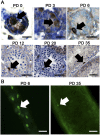
Identification of Id4-Gfp-expressing spermatogonia in testes of LT-11B6 mice at multiple stages of postnatal life. (A) Representative images of immunostaining for Id4-Gfp-expressing cells in cross-sections of seminiferous tubules from testes of mice at PD 0, 3, 6, 12, 20, and 35. Arrows indicate Id4-Gfp+ spermatogonia. Bars: PD 0, 3, and 6, 50 μm; PD 12, 20, and 35, 100 μm. (B) Representative pictures of live imaging for Id4-Gfp+ cells in whole-mount seminiferous tubules from LT11-B6 mice at PD 6 and 35. Arrows indicate single Id4-Gfp+ spermatogonia. Bars, 50 μm.
Figure 3.
Figure 3.
Proportionality of Id4-Gfp-expressing cells in the undifferentiated spermatogonial population of testes from LT-11B6 mice at multiple stages of postnatal life. (A) Representative images of coimmunofluorescent staining for Plzf (red) and Id4-Gfp-expressing cells in cross-sections of seminiferous tubules from testes of mice at PD 3, 6, 12, 20, and 35. DAPI (blue) was used to stain the nuclei of cells. At each age, the right image is a magnified view of the left image. Arrows indicate Plzf+/Id4-Gfp+ spermatogonia, and stars indicate Plzf+/Id4-Gfp spermatogonia. Note that four images are provided for PD 35 to demonstrate multiple different spermatogonia. Bars: left images, 100 μm; right images, 50 μm. (B) Quantification of the proportion of the undifferentiated spermatogonial population that is Id4-Gfp+ at multiple stages of postnatal development. Data are presented as mean ± SEM percentage of the Plzf+ undifferentiated spermatogonial population and were generated from immunostained cross-sections of three different mice at each age point. (C) Representative image of coimmunofluorescent staining for spermatogonia expressing the markers Gfra1 (red) and Id4-Gfp (green) within whole-mount seminiferous tubules from mice at PD 6. DAPI (blue) was used to stain the nuclei of cells. Arrows indicate single spermatogonia that are Gfra1+/Id4-Gfp+, and stars indicate spermatogonia that are Gfra1+/Id4-Gfp. Bar, 100 μm.
Figure 4.
Figure 4.
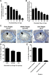
Frequency and distribution of Id4-Gfp-expressing spermatogonia in testes of LT-11B6 mice during postnatal development and adulthood. (A) Quantification of seminiferous tubule cross-sections from testes of mice at PD 0, 3, 6, 12, 20, and 35 containing at least one Id4-Gfp+ spermatogonia. Data are presented as the mean ± SEM percentage of total seminiferous tubule cross-sections. (B) Quantification of Id4-Gfp+ spermatogonial numbers in cross-sections of seminiferous tubules from testes of mice at PD 0, 3, 6, 12, 20, and 35. Data are presented as the mean ± SEM number of positive cells per seminiferous tubule cross-section. (C) Representative images of immunostaining for Id4-Gfp-expressing cells in cross-sections of seminiferous tubules from testes of adult mice grouped as early (II–VI), middle (VII–VIII), or late (IX–I) stages of the seminiferous epithelial cycle. Arrows indicate Id4-Gfp+ spermatogonia. Bars, 50 μm. (D) Quantification of seminiferous tubule cross-sections at early, middle, and late stages of the seminiferous epithelial cycle containing at least one Id4-Gfp+ spermatogonia. Data are presented as mean ± SEM percentage of the total Id4-Gfp+ spermatogonial population. (E) Quantification of regions within seminiferous tubule cross-sections where Id4-Gfp+ spermatogonia reside. Regions were defined as being juxtaposed to other tubules, interstitial tissue not containing blood vessels (−BV), and interstitial tissue containing blood vessels (+BV). Data are presented as mean ± SEM percentage of the total Id4-Gfp+ spermatogonial population.
Figure 5.
Figure 5.
Regenerative capacity of Id4-Gfp+ and Id4-Gfp spermatogonial populations from testes of hybrid LT-11B6/RosaLacZ mice. (A) Representative image of overlaid fluorescent and phase-contrast views of primary cultures of undifferentiated spermatogonia established from LT-11B6/RosaLacZ mice. Black arrows indicate clumps of spermatogonia containing Id4-Gfp+ cells, and white arrows indicate clumps not containing Id4-Gfp+ cells. Note that all spermatogonia (i.e., Gfp+ and Gfp) express LacZ. (B) Representative post-sort scatter plots of Id4-Gfp+ and Id4-Gfp populations isolated from primary cultures of undifferentiated spermatogonia using fluorescence-activated cell sorting (FACS). (C) Representative images of testes from recipient mice 2 mo after transplantation with FACS-isolated Id4-Gfp+ and Id4-Gfp subpopulations that have been stained for LacZ-expressing colonies of donor-derived spermatogenesis. Note that each blue segment is clonally derived from a single SSC. (D) Quantitative comparison of donor-derived colonies of spermatogenesis in testes of recipient mice that were generated from 105 FACS-isolated Id4-Gfp+ or Id4-Gfp cells 2 mo after transplantation. Data are mean ± SEM for three different cultures. (*)Significantly different at P < 0.05.
Figure 6.
Figure 6.
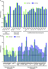
Transcriptome analysis of Id4-Gfp+ and Id4-Gfp spermatogonial populations from testes of LT-11B6 mice. (A) Quantitative comparison of transcript abundance for genes involved in specification of primordial germ cells (PGCs) and germ cell licensing and several genes reported previously to be expressed by undifferentiated spermatogonia. (B) Quantitative comparison of transcripts with significantly (Q < 0.05) different abundance. For both data sets, Id4-Gfp+ and Id4-Gfp fractions were isolated from primary cultures of undifferentiated spermatogonia, and transcript abundances were determined using RNA-seq analysis. Data are presented as log10 of the mean FPKM generated from three different cultures. Differential abundance in B was determined statistically using Cuffdiff analysis. Note that dashed lines in A and B are set at 1, which was considered the cutoff of a transcript being considered present or absent.
Figure 7.
Figure 7.
The hierarchy for subsets of spermatogonia in the undifferentiated population of mouse testes and expression profile for intracellular molecular markers. The Asingle spermatogonial pool consists of SSC and transitional progenitor subtypes. The SSC subtype possesses regenerative capacity and unique self-renewal properties to remain as Asingle following division, thereby sustaining a pool from which new transitional Asingle progenitor spermatogonia arise. In contrast, the transitional subtype attains enhanced propensity to form initial chained spermatogonia (Apaired) upon the next division and potentially retains limited capacity for self-renewal but lacks regenerative capacity. The SSC and transitional Asingle subtypes express genes that are also expressed by chained (Apaired-aligned) progenitors but are distinguished by expression of specific genes such as Id4 and Utf1. Gene expression that distinguishes the transitional Asingle progenitor spermatogonia is undefined but may include reduced levels of some genes also expressed by Asingle SSCs.

References

    1. Arnold K, Sarkar A, Yram MA, Polo JM, Bronson R, Sengupta S, Seandel M, Geijsen N, Hochedlinger K 2011. Sox2(+) adult stem and progenitor cells are important for tissue regeneration and survival of mice. Cell Stem Cell 9: 317–329 - PMC - PubMed
    1. Barker N, van Es JH, Kuipers J, Kujala P, van den Born M, Cozijnsen M, Haegebarth A, Korving J, Begthel H, Peters PJ, et al. 2007. Identification of stem cells in small intestine and colon by marker gene Lgr5. Nature 449: 1003–1007 - PubMed
    1. Bentley KL, Shashikant CS, Wang W, Ruddle NH, Ruddle FH 2010. A yeast-based recombinogenic targeting toolset for transgenic analysis of human disease genes. Ann N Y Acad Sci 1207: E58–E68 - PubMed
    1. Brinster RL 2002. Germline stem cell transplantation and transgenesis. Science 296: 2174–2176 - PMC - PubMed
    1. Brinster RL 2007. Male germline stem cells: from mice to men. Science 316: 404–405 - PMC - PubMed

Publication types

MeSH terms

Substances

Associated data