Review
. 2014 May 22:3:12.
doi: 10.1186/2001-1326-3-12. eCollection 2014.

Pluripotent muse cells derived from human adipose tissue: a new perspective on regenerative medicine and cell therapy

Affiliations
Review

Pluripotent muse cells derived from human adipose tissue: a new perspective on regenerative medicine and cell therapy

Ariel A Simerman et al. Clin Transl Med. .

Abstract

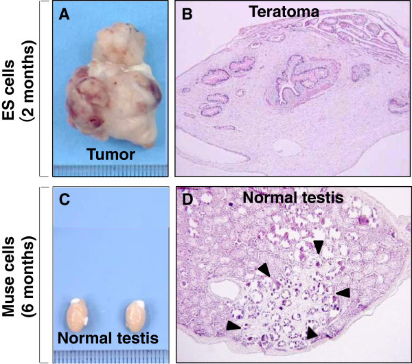
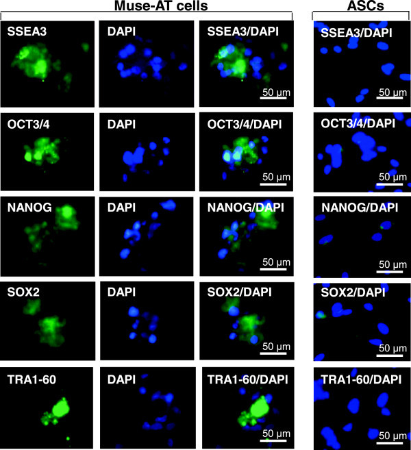
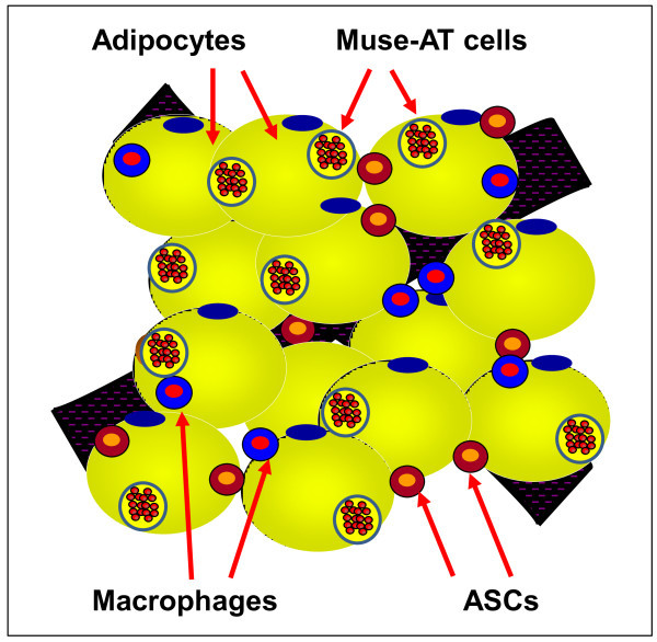
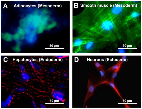
In 2010, Multilineage Differentiating Stress Enduring (Muse) cells were introduced to the scientific community, offering potential resolution to the issue of teratoma formation that plagues both embryonic stem (ES) and induced pluripotent (iPS) stem cells. Isolated from human bone marrow, dermal fibroblasts, adipose tissue and commercially available adipose stem cells (ASCs) under severe cellular stress conditions, Muse cells self-renew in a controlled manner and do not form teratomas when injected into immune-deficient mice. Furthermore, Muse cells express classic pluripotency markers and differentiate into cells from the three embryonic germ layers both spontaneously and under media-specific induction. When transplanted in vivo, Muse cells contribute to tissue generation and repair. This review delves into the aspects of Muse cells that set them apart from ES, iPS, and various reported adult pluripotent stem cell lines, with specific emphasis on Muse cells derived from adipose tissue (Muse-AT), and their potential to revolutionize the field of regenerative medicine and stem cell therapy.

Keywords: Adult pluripotent stem cells; Muse cells; Non-tumorigenic; Quiescence; Regenerative medicine.


Figures

Figure 1
Figure 1
Non-tumorigenicity of Muse cells. (A) Embryonic stem (ES) cells infused into immunodeficient mice (SCID mice) testes, formed teratomas within 8 to 12 weeks, (B) Histological analysis showed that the teratoma contained muscle tissue, intestine-like structures and keratinized skin, (C) Muse cell-transplanted testes did not generate teratomas similar to untreated testes, and (D) Testes injected with Muse cells maintained normal structure even 6 months post-injection (Pictures reproduced from Proc Natl Acad Sci USA 2010, 107: 8639–43, and Stem Cells Dev 2014 DOI: 10.1089/scd.2013.0473).
Figure 2
Figure 2
Muse-ATs express pluripotent stem cell markers. Immunofluorescence microscopy demonstrates that Muse-AT aggregates, along with individual Muse-AT cells, express characteristic pluripotent stem cell markers, including SSEA3, Oct3/4, Nanog, Sox2, and TRA-1-60 [24,26] (Figure 2). Comparatively, ASCs (right panel) derived from the same lipoaspirate under standard conditions [38] were negative for these pluripotent stem cell markers. Nuclei were stained with DAPI (blue). Original magnification, 600 X (Pictures reproduced from PLoS One 2013, 8(6):e64752).
Figure 3
Figure 3
Graphical depiction of different cell components present in adipose tissue. Adipose tissue is composed of adipocytes and the stromal vascular fraction containing adipose tissue macrophages (ATMs), adipose stem cells (ASCs) and Muse-AT cells, among other cell components.
Figure 4
Figure 4
Tripoblastic characteristics of Muse-AT cells. Muse-AT cells were grown as adherent cells in the presence of (A) adipogenic medium; the formation of adipocytes was detected using BODYI-PI-C16 which identify lipid drops present in adipocytes; (B) myogenic differentiation medium; the formation of myocytes was detected using an anti-human MSA antibody; (C) hepatogenic differentiation medium; formation of hepatocytes was detected using an anti-cytokeratin 7 antibody; (D) Muse-AT cells were grown for 7 days as non-adherent cells and then cultured for an additional 7 days as adherent cells; neural-like cells were detected by immunofluorescence using an anti-human MAP2 antibody. Nuclei were stained with DAPI (blue). Original magnification was 600X. (Pictures reproduced from PLoS One 2013, 8(6):e64752).

References

    1. Evans MJ, Kaufman MH. Establishment in culture of pluripotential cells from mouse embryos. Nature. 1981;3(5819):154–156. doi: 10.1038/292154a0. - DOI - PubMed
    1. Martin GR. Isolation of a pluripotent cell line from early mouse embryos cultured in medium conditioned by teratocarcinoma stem cells. Proc Natl Acad Sci U S A. 1981;3(12):7634–7638. doi: 10.1073/pnas.78.12.7634. - DOI - PMC - PubMed
    1. Thomson JA, Itskovitz-Eldor J, Shapiro SS, Waknitz MA, Swiergiel JJ, Marshall VS, Jones JM. Embryonic stem cell lines derived from human blastocysts. Science. 1998;3(5391):1145–1147. - PubMed
    1. Przyborski SA. Differentiation of human embryonic stem cells after transplantation in immune-deficient mice. Stem Cells. 2005;3(9):1242–1250. doi: 10.1634/stemcells.2005-0014. - DOI - PubMed
    1. Maherali N, Sridharan R, Xie W, Utikal J, Eminli S, Arnold K, Stadtfeld M, Yachechko R, Tchieu J, Jaenisch R, Plath K, Hochedlinger K. Directly reprogrammed fibroblasts show global epigenetic remodeling and widespread tissue contribution. Cell Stem Cell. 2007;3(1):55–70. doi: 10.1016/j.stem.2007.05.014. - DOI - PubMed