. 2014 Jun 1;3(6):405-413.
doi: 10.1089/wound.2014.0539.

The Effect of Bone-Marrow-Derived Stem Cells and Adipose-Derived Stem Cells on Wound Contraction and Epithelization

Affiliations

The Effect of Bone-Marrow-Derived Stem Cells and Adipose-Derived Stem Cells on Wound Contraction and Epithelization

Cagri A Uysal et al. Adv Wound Care (New Rochelle). .

Abstract

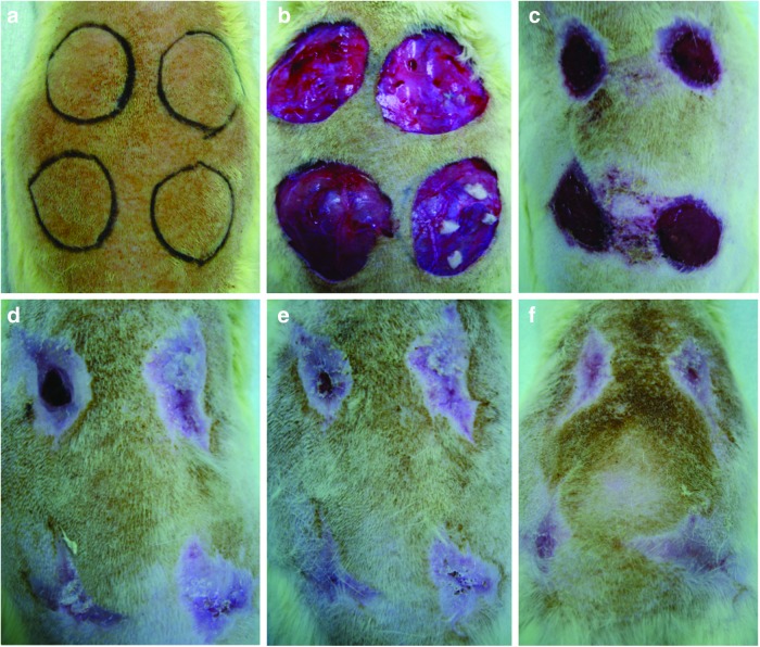
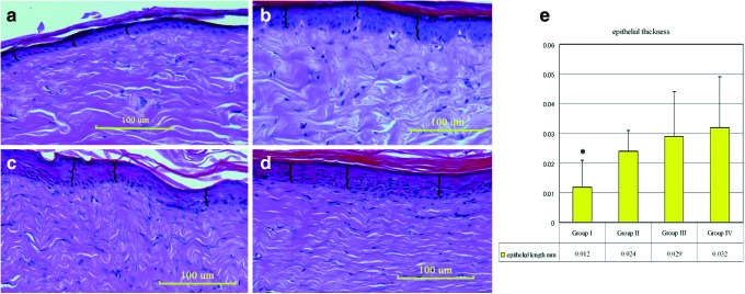
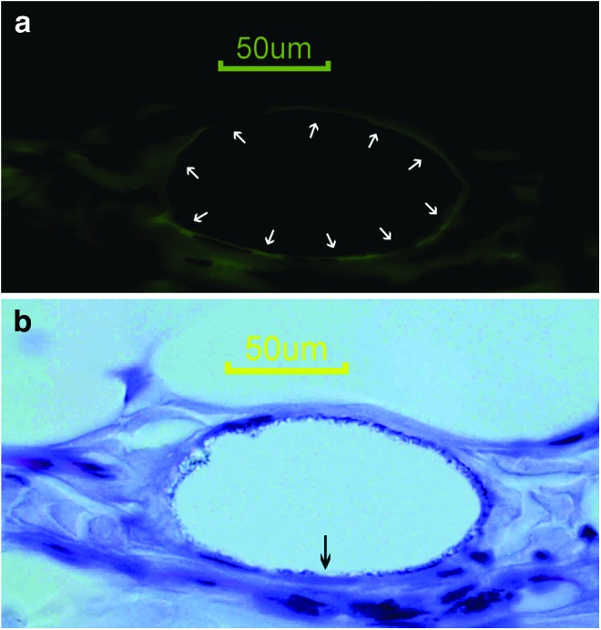
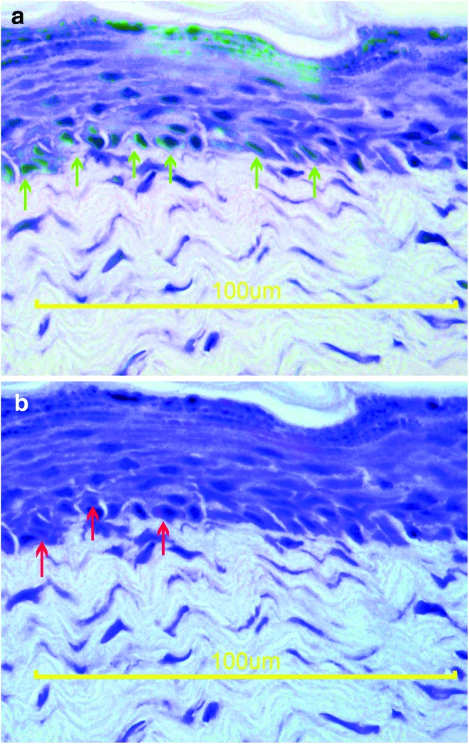
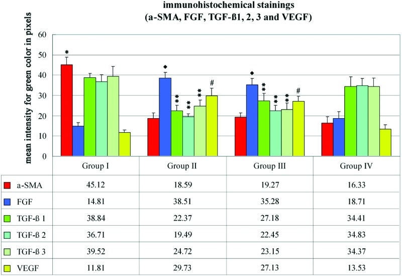
Objective: The relationship between the wound contraction and levels of α-smooth muscle actin (α-SMA) has been revealed in different studies. We aimed to investigate the effects of mesenchymal stem cells (MSCs), mainly bone-marrow-derived stem cells (BSCs) and adipose-derived stem cells (ASCs), and find out the α-SMA, fibroblast growth factor (FGF), transforming growth factor beta, and vascular endothelial growth factor (VEGF) levels on an in vivo acute wound healing model after the application of MSCs. Approach: Four circular skin defects were formed on the dorsum of Fisher rats (n=20). The defects were applied phosphate-buffered saline (PBS), ASCs, BSCs, and patchy skin graft, respectively. The healing time and scar area were noted. Results: There was a statistical decrease in the healing time in ASC, BSC, and skin graft groups (p<0.05). However, the scar was smaller in the PBS group (p<0.05). The α-SMA levels were statistically lower in ASC, BSC, and graft groups (p<0.05). The FGF levels were statistically higher in ASC and BSC groups (p<0.05). The differentiation of the injected MSCs to endothelial cells and keratinocytes was observed. Innovation and Conclusion: MSCs decrease the healing time and contraction of the wound while increasing the epithelization rate by increasing angiogenesis.


Figures

None
Cagri A. Uysal, MD, PhD
<b>Figure 1.</b>
Figure 1.
(a) Four circular skin defects with a diameter of 20 mm were planned on the dorsum of the rat. (b) Group I (left cranial): 1 mL phosphate-buffered saline (PBS) injection; Group II (right cranial): 1 mL PBS mixed with 1×10⁷ ASCs; Group III (left caudal): 1 mL PBS mixed with 1×10⁷ BSCs; Group IV (right caudal): patchy skin grafts following 1 mL PBS injection. Wound healing progress was photographed on postoperative days 21 (c), 42 (d), 49 (e), and 56 (f). To see this illustration in color, the reader is referred to the web version of this article at www.liebertpub.com/wound
<b>Figure 2.</b>
Figure 2.
Photographs indicate the hematoxylin and eosin staining of the specimens: groups I (a), II (b), III (c), and IV (d). Braces “{” indicate the length between the stratum basale and stratum corneum calculated in 20 different fields under×40 magnification (scale bar=100 μm). (e) Graph describing the mean epithelial thickness. There was a statistical difference between group I and groups II, III, and IV (*p<0.05). To see this illustration in color, the reader is referred to the web version of this article at www.liebertpub.com/wound
<b>Figure 3.</b>
Figure 3.
(a) Anti-vWF antibody immunohistochemical staining reveals the endothelial lining (white arrows). (b) The hematoxylin staining and DiI fluorescence photographs were merged to localize the DiI positivity. The black arrow indicates a DiI-positive endothelial cell (scale bar=50 μm). To see this illustration in color, the reader is referred to the web version of this article at www.liebertpub.com/wound
<b>Figure 4.</b>
Figure 4.
(a) The hematoxylin and anti-cytokeratin antibody immunohistochemical stainings were merged to localize the positivity. The green arrows indicate the keratinocytes at the basal membrane. (b) The hematoxylin staining and DiI fluorescence photographs were merged to localize the DiI positivity. The red arrows indicate keratinocytes originating from the injected stem cell (scale bar=100 μm). To see this illustration in color, the reader is referred to the web version of this article at www.liebertpub.com/wound
<b>Figure 5.</b>
Figure 5.
The immunohistochemical staining graph for alpha smooth muscle actin (α-SMA); fibroblast growth factor (FGF); transforming growth factor (TGF)-β1, -β2, and -β3; and vascular endothelial growth factor (VEGF). The anti–α-SMA antibody levels in groups II, III, and IV were statistically lower than group I (*p<0.05). The anti-FGF levels in groups II and III were statistically higher when compared with groups I and IV (p<0.05). The anti–TGF-β subfamily antibody levels were statistically lower in groups II and III (**p<0.05). The VEGF levels were statistically higher in groups II and III when compared with groups I and IV (#p<0.05). To see this illustration in color, the reader is referred to the web version of this article at www.liebertpub.com/wound

References

    1. Williams IR. and Kupper TS: Immunity at the surface: homeostatic mechanisms of the skin immune system. Life Sci 1996; 58:1485. - PubMed
    1. Desmouliere A. and Gabbiani G: Modulation of fibroblastic cytoskeletal features during pathological situations: the role of extracellular matrix and cytokines. Cell Motil Cytoskeleton 1994; 29:195. - PubMed
    1. Gabbiani G, Ryan GB, and Majne G: Presence of modified fibroblasts in granulation tissue and their possible role in wound contraction. Experientia 1971; 27:549. - PubMed
    1. Desmouliere A. and Gabbiani G: The role of myofibroblast in wound healing and fibrocontractive diseases. In: The Molecular and Cellular Biology of Wound Repair, 2nd edition, edited by Clark RAF. New York, Plenum, 1996, pp. 321–323
    1. Desmouliere A, Chaponnier C, and Gabbiani G: Tissue repair, contraction, and the myofibroblast. Wound Repair Regen 2005; 13:7. - PubMed

LinkOut - more resources