. 2014 Jun 18;9(6):e98781.
doi: 10.1371/journal.pone.0098781. eCollection 2014.

Foldscope: origami-based paper microscope

Affiliations

Foldscope: origami-based paper microscope

James S Cybulski et al. PLoS One. .

Abstract

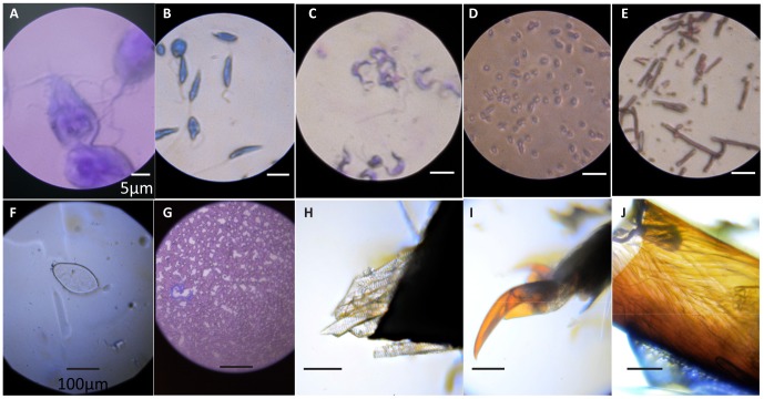
Here we describe an ultra-low-cost origami-based approach for large-scale manufacturing of microscopes, specifically demonstrating brightfield, darkfield, and fluorescence microscopes. Merging principles of optical design with origami enables high-volume fabrication of microscopes from 2D media. Flexure mechanisms created via folding enable a flat compact design. Structural loops in folded paper provide kinematic constraints as a means for passive self-alignment. This light, rugged instrument can survive harsh field conditions while providing a diversity of imaging capabilities, thus serving wide-ranging applications for cost-effective, portable microscopes in science and education.


Conflict of interest statement

Competing Interests: This work is covered under patent application PCT/US2013/025612 (Title: Optical Device). Further details, including the 10,000 microscope project and updates on the latest status of this work, are available on www.foldscope.com. This does not alter the authors' adherence to PLOS ONE policies on sharing data and materials.

Figures

Figure 1
Figure 1. Foldscope design, components and usage.
(A) CAD layout of Foldscope paper components on an A4 sheet. (B) Schematic of an assembled Foldscope illustrating panning, and (C) cross-sectional view illustrating flexure-based focusing. (D) Foldscope components and tools used in the assembly, including Foldscope paper components, ball lens, button-cell battery, surface-mounted LED, switch, copper tape and polymeric filters. (E) Different modalities assembled from colored paper stock. (F) Novice users demonstrating the technique for using the Foldscope. (G) Demonstration of the field-rugged design, such as stomping under foot.
Figure 2
Figure 2. Foldscope imaging modalities.
(A) Brightfield Foldscope image of a monolayer of 1 µm polystyrene microspheres (Polysciences 07310-15) using a 1,450X lens. (B) Fluorescent Foldscope image of 2 µm polyfluorescent microspheres (Polysciences 19508-2) using a 1,140X lens with Roscolux gel filters #19 and #80. (C) 2X2 lens-array Brightfield Foldscope image of Giemsa-stained thin blood smear using 1,450X lenses. (D) 140X Darkfield Foldscope images of 6 µm polystyrene microspheres (Polysciences 15714-5), using a 140X lens for the darkfield condenser. Darkfield condenser aperture shown in inset has 1.5 mm inner diameter and 4.0 mm outer diameter. (E–H) Schematic cross-sections of Brightfield, Fluorescence, Lens-Array, and Darkfield Foldscope configurations, showing the respective arrangements of ball lenses, filters, and LEDs. See table 2 for ball lenses used for specific magnifications.
Figure 3
Figure 3. Manufacturing innovations for lens- and specimen- mounting.
(A) Fabrication, mounting, and characterization of capillary-encapsulation process for lens-mounted apertures. X and Y error bars for all measurements are 2.5 µm. (B) Reel of polystyrene carrier tape with custom pockets and punched holes for mounting over 2,000 ball lenses with optimal apertures. The first ten pockets include mounted ball lenses. Inset shows sectioned view from CAD model of carrier tape mounted lenses. Note the aperture is the punched hole shown on the bottom side of the ball lens. This tape is 16 mm wide and is designed for 2.4 mm ball lenses (aperture diameter is 0.7 mm). (C) Top: Paper microscope slide shown next to standard glass slide with coverslip, both with wet mount algae specimens. Bottom: Schematic of paper microscope slide, showing specimen containment cavity formed between upper tape and lower tape in middle of slide.
Figure 4
Figure 4. Analytical, numerical, and empirical characterization of Foldscope.
(A,B) Analytical “design curves” for normalized optimal aperture radius (nOAR) and optimal resolution (RES) versus magnification (MAG) over index of refraction (range 1.33–1.91) and ball lens radius (range 40–1200 µm). (C) Comparison of analytical (3D surface) and numerical (plotted as points) results for RES versus index of refraction and ball lens radius. (D) Modulus of the Optical Transfer Function (MTF) over the optimal field of view for a 300 µm sapphire lens with optimal aperture. (E,F) Image of USAF 1951 resolution target taken with 430X ball lens, including an enlarged caption of Group 9, and an intensity profile plot along path denoted by green line in image caption. This demonstrates resolvability for Group 9, Element 4 corresponding to 724 Line Pairs/mm or 1.38 µm resolution. (G,H) Image of USAF 1951 resolution target taken with 140X ball lens, including an enlarged caption of Group 8, and an intensity profile plot along path denoted by green line in image caption. This demonstrates resolvability for Group 8, Element 6 corresponding to 456 Line Pairs/mm or 2.19 µm resolution. The data was taken using GUPPY Pro 503C scientific camera, with 2592×1944 pixels and pixel size 2.2×2.2 µm2.
Figure 5
Figure 5. Mosaic of Foldscope Images.
Bright field images of (A) Giardia lamblia (2,180X), (B) Leishmania donovani (1,450X), (C) Trypanosoma cruzi (1,450X), (D) gram-negative Escherichia coli (1,450X), (E) gram-positive Bacillus cereus (1,450X), (F) Schistosoma haematobium (140X), and (G) Dirofilaria immitis (140X). Unstained (H) leg muscles and (I) tarsi of an unidentified ladybug (genus Coccinella). (J) Unstained leg muscles (fixed in formaldehyde) of an unidentified red ant (genus Solenopsis). An LED diffuser (Roscolux #111) was added for (A) and an LED condenser (2.4 mm borosilicate ball lens) was used for (C). Images (H–J) were taken by novice user using a self-made Foldscope (140X). See table 2 for ball lenses used for specific magnifications. White scale bar: 5 µm; black scale bar: 100 µm.

References

    1. Keller E, Goldman R (2006) Light Microscopy. Woodbury, NY: Cold Spring Harbor Laboratory Press. p. 8.
    1. Whitesides GM (2011) The Frugal Way. The Economist - The World in 2012
    1. Rothemund PW (2006) Folding DNA to create nanoscale shapes and patterns. Nature 440: 297–302. - PubMed
    1. Hyde RA (1999) Eyeglass, a large-aperture space telescope. Appl Opt 38 19: 4198–4212. - PubMed
    1. Hoover AM, Fearing RS (2008) Fast scale prototyping for folded millirobots. IEEE International Conference on Robotics and Automation 5: 886–892.

Publication types