. 2014 Jun 17;106(12):2625-35.
doi: 10.1016/j.bpj.2014.04.046.

Lateral motion and bending of microtubules studied with a new single-filament tracking routine in living cells

Affiliations

Lateral motion and bending of microtubules studied with a new single-filament tracking routine in living cells

Carla Pallavicini et al. Biophys J. .

Abstract

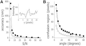
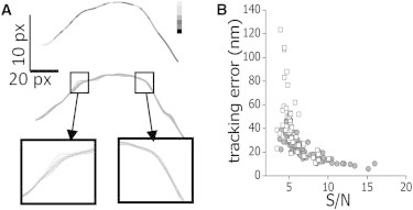
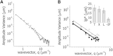
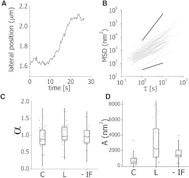
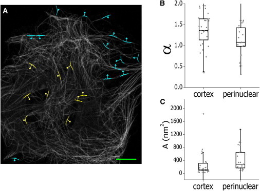
The cytoskeleton is involved in numerous cellular processes such as migration, division, and contraction and provides the tracks for transport driven by molecular motors. Therefore, it is very important to quantify the mechanical behavior of the cytoskeletal filaments to get a better insight into cell mechanics and organization. It has been demonstrated that relevant mechanical properties of microtubules can be extracted from the analysis of their motion and shape fluctuations. However, tracking individual filaments in living cells is extremely complex due, for example, to the high and heterogeneous background. We introduce a believed new tracking algorithm that allows recovering the coordinates of fluorescent microtubules with ∼9 nm precision in in vitro conditions. To illustrate potential applications of this algorithm, we studied the curvature distributions of fluorescent microtubules in living cells. By performing a Fourier analysis of the microtubule shapes, we found that the curvatures followed a thermal-like distribution as previously reported with an effective persistence length of ∼20 μm, a value significantly smaller than that measured in vitro. We also verified that the microtubule-associated protein XTP or the depolymerization of the actin network do not affect this value; however, the disruption of intermediate filaments decreased the persistence length. Also, we recovered trajectories of microtubule segments in actin or intermediate filament-depleted cells, and observed a significant increase of their motion with respect to untreated cells showing that these filaments contribute to the overall organization of the microtubule network. Moreover, the analysis of trajectories of microtubule segments in untreated cells showed that these filaments presented a slower but more directional motion in the cortex with respect to the perinuclear region, and suggests that the tracking routine would allow mapping the microtubule dynamical organization in cells.


Figures

Figure 1
Figure 1
Motion of microtubules in living cells. (A) Dynamics of microtubules in a small region of the cell (yellow square in left panel); the time lag between frames was 60 s. (Yellow star) Fixed reference point. Scale bar, 10 μm. (B) Intensity profiles recovered along the yellow lines for the microtubules observed in the zoom-in images. The S/N of these filaments was ∼10. To see this figure in color, go online.
Figure 2
Figure 2
Performance of the filament-tracking routine. (A) We simulated 20 confocal images of a single, fluorescent filament and used the proposed neural network-based tracking routine (○) or a Gaussian deconvolution-based algorithm (■) to track its position. The error on the filament position determination was calculated using Eq. 5. (Inset) A representative intensity profile obtained perpendicular to a microtubule labeled with EGFP-XTP (S/N = 5) in X. laevis melanophores was fitted using the neural network-based tracking routine (continuous line) or a Gaussian function (dotted lines). (B) Twenty confocal images of two intersecting filaments (S/N = 10) were simulated and tracked using the neural network-based tracking routine (○) or a Gaussian deconvolution-based algorithm (■). The confusion regions predicted according to the diffraction limit (▵) and those obtained with the tracking routines are plotted as a function of the intersecting angle.
Figure 3
Figure 3
Tracking precision. Rhodamine-labeled microtubules adsorbed onto a coverslip were imaged as a function of time (frame time = 3.3 s) following the sequence of photobleaching and imaging described in the text. The image stacks were analyzed with the filament-tracking routine and the tracking error was determined for microtubules presenting different S/N values as described in the text. (A) Pseudocolor image of a representative microtubule showing the intensity recovered at each position (from black to white, white representing high intensities) (top panel). Recovered shapes of the microtubule in 20 consecutive frames (middle panels). Zoom-in of the shapes recovered in regions of the microtubule presenting low and high intensities (bottom panels). (B) Tracking error for fixed microtubules (●) and of EGFP-XTP-labeled microtubules in formaldehyde-fixed melanophore cells (□) as a function of the signal/noise. The pixel size was 100 nm.
Figure 4
Figure 4
Fourier analysis of microtubule shapes. (A) Images of 8 μm-long filaments (N = 50) with shapes following a thermal distribution with lp = 15 μm were simulated as described in the Supporting Material setting S/N = 10. Equation 6 was fitted to the simulated data in the range 1< n < 9 (continuous line) obtaining lp = 16 ± 3 μm. (B) The variance of the Fourier mode amplitudes of microtubules was determined for cells transfected with EGFP α-tubulin (■, T), EGFP-XTP cells (▼, X), and EGFP-XTP cells treated with latrunculin to disrupt the actin filament network (○, L) or EGFP-XTP cells transfected with a dominant-negative construct of EGFP-vimentin that disrupts the intermediate-filament network (▵, −IF). (Solid lines) Least-squares fit to a thermal-like behavior (Eq. 6). The figure includes data from 80 to 110 filaments and from 15 to 50 cells in each experimental condition. (Inset) Apparent persistence lengths obtained in the assayed conditions.
Figure 5
Figure 5
Microtubule lateral motion. (A) Representative time course plot of the average lateral position (LP) of a microtubule segment. (B) MLSD data (Eq. 4) as a function of the time lag obtained for 96 filaments. (Black lines) Representative behavior expected for subdiffusion with α = 0.5 (bottom) and ballistic motion (α = 2, top). (C and D) The MLSD data were fitted using the anomalous diffusion model (Eq. 7). Box plots of the anomalous diffusion coefficient α (C) and preexponential factor A (D) obtained for control (C), latrunculin-treated (L), and vimentin-disrupted (−IF) EGFP-XTP cells. The figure includes data from 80 to 110 filaments and from 15 to 50 cells in each experimental condition.
Figure 6
Figure 6
Microtubules motion depends on their relative position within the cell. (A) (Grayscale image) EGFP-XTP cell showing some tracked filament segments in the perinuclear region (circleheads) or near the cortex (arrowheads). (Lines) Vectors defined by the initial and final positions determined for each segment. Scale bar, 10 μm. The MLSD data were fitted using the anomalous diffusion model (Eq. 7). Box plots of the anomalous diffusion exponent α (B) and preexponential factor A (C) obtained by fitting Eq. 7. The figure includes data from 51 filaments from five different cells. To see this figure in color, go online.

References

    1. Fletcher D.A., Mullins R.D. Cell mechanics and the cytoskeleton. Nature. 2010;463:485–492. - PMC - PubMed
    1. Mouneimne G., Hansen S.D., Brugge J.S. Differential remodeling of actin cytoskeleton architecture by profilin isoforms leads to distinct effects on cell migration and invasion. Cancer Cell. 2012;22:615–630. - PMC - PubMed
    1. Goldstein D., Elhanan T., Weihs D. Origin of active transport in breast-cancer cells. Soft Matter. 2013;9:7167–7173.
    1. Howard J. Sinauer Associates; Sunderland, MA: 2001. Mechanics of Motor Proteins and the Cytoskeleton.
    1. Mitchison T., Kirschner M. Dynamic instability of microtubule growth. Nature. 1984;312:237–242. - PubMed

Publication types

LinkOut - more resources