. 2014 Jun 18;9(6):e99988.
doi: 10.1371/journal.pone.0099988. eCollection 2014.

DMXAA causes tumor site-specific vascular disruption in murine non-small cell lung cancer, and like the endogenous non-canonical cyclic dinucleotide STING agonist, 2'3'-cGAMP, induces M2 macrophage repolarization

Affiliations

DMXAA causes tumor site-specific vascular disruption in murine non-small cell lung cancer, and like the endogenous non-canonical cyclic dinucleotide STING agonist, 2'3'-cGAMP, induces M2 macrophage repolarization

Charlene M Downey et al. PLoS One. .

Abstract

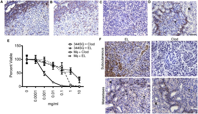
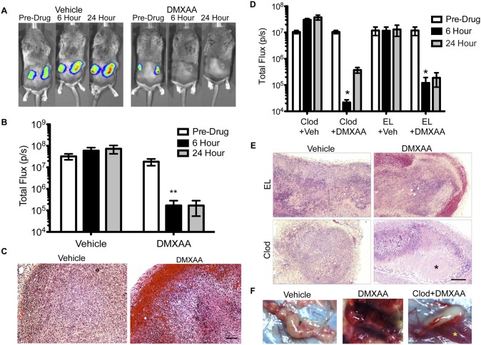
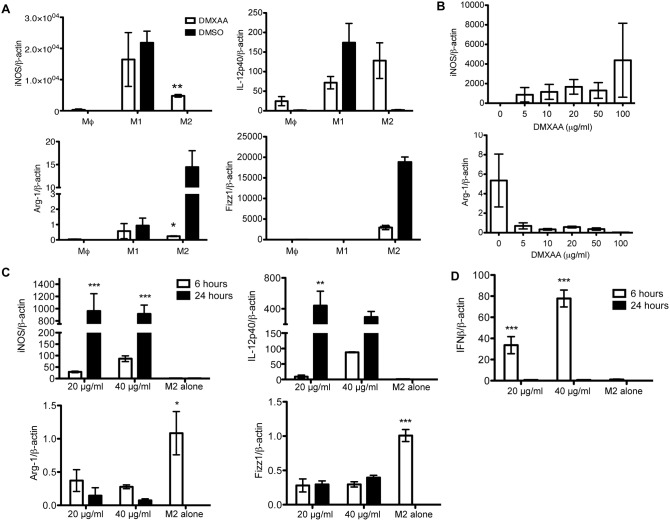
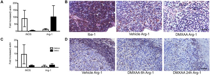
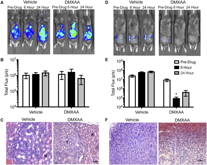
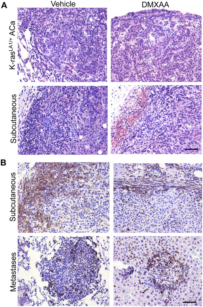
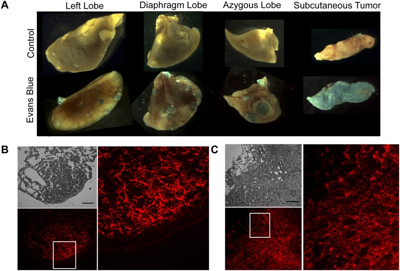
The vascular disrupting agent 5,6-dimethylxanthenone-4-acetic acid (DMXAA), a murine agonist of the stimulator of interferon genes (STING), appears to target the tumor vasculature primarily as a result of stimulating pro-inflammatory cytokine production from tumor-associated macrophages (TAMs). Since there were relatively few reports of DMXAA effects in genetically-engineered mutant mice (GEMM), and models of non-small cell lung cancer (NSCLC) in particular, we examined both the effectiveness and macrophage dependence of DMXAA in various NSCLC models. The DMXAA responses of primary adenocarcinomas in K-rasLA1/+ transgenic mice, as well as syngeneic subcutaneous and metastatic tumors, generated by a p53R172HΔg/+; K-rasLA1/+ NSCLC line (344SQ-ELuc), were assessed both by in vivo bioluminescence imaging as well as by histopathology. Macrophage-dependence of DMXAA effects was explored by clodronate liposome-mediated TAM depletion. Furthermore, a comparison of the vascular structure between subcutaneous tumors and metastases was carried out using micro-computed tomography (micro-CT). Interestingly, in contrast to the characteristic hemorrhagic necrosis produced by DMXAA in 344SQ-ELuc subcutaneous tumors, this agent failed to cause hemorrhagic necrosis of either 344SQ-ELuc-derived metastases or autochthonous K-rasLA1/+ NSCLCs. In addition, we found that clodronate liposome-mediated depletion of TAMs in 344SQ-ELuc subcutaneous tumors led to non-hemorrhagic necrosis due to tumor feeding-vessel occlusion. Since NSCLC were comprised exclusively of TAMs with anti-inflammatory M2-like phenotype, the ability of DMXAA to re-educate M2-polarized macrophages was examined. Using various macrophage phenotypic markers, we found that the STING agonists, DMXAA and the non-canonical endogenous cyclic dinucleotide, 2'3'-cGAMP, were both capable of re-educating M2 cells towards an M1 phenotype. Our findings demonstrate that the choice of preclinical model and the anatomical site of a tumor can determine the vascular disrupting effectiveness of DMXAA, and they also support the idea of STING agonists having therapeutic utility as TAM repolarizing agents.


Conflict of interest statement

Competing Interests: The authors have declared that no competing interests exist.

Figures

Figure 1
Figure 1. Effectiveness of clodronate-mediated TAM depletion varied depending on tumor site.
Representative 344SQ-ELuc subcutaneous tumor sections were stained with antibodies against Iba-1 (A) and Arg-1 (B), demonstrating abundant M2-like macrophages primarily at the tumor periphery. In contrast, much lower and variable macrophage infiltration was present in either the K-rasLA1/+ primary NSCLCs (C) or in 344SQ-ELuc metastases (D). (E) MTT assay conducted on BMDM (Mφ) or 344SQ-ELuc cells to assess potential cytotoxicity in response to either Clodrolip (Clod) or empty liposomes (EL). (F) Representative 344SQ-ELuc subcutaneous tumor and kidney metastases sections from Clod-treated mice were stained with Iba-1 showing that TAM depletion only occurred in subcutaneous tumors (T = tumor, K = kidney). Scale bar = 100 µm (A–B) 50 µm (C, D, F). Data represent the mean ± SEM.
Figure 2
Figure 2. TAM depletion prevented DMXAA-induced hemorrhagic necrosis of subcutaneous NSCLC.
(A) BLI of bilateral 344SQ-ELuc subcutaneous tumors in a representative syngeneic 129/Sv mouse at day 14 post-cell injection. Mice were randomized into two groups and administered a single i.p. dose of 25 mg/kg DMXAA or DMSO vehicle and imaged again at 6 and 24 hours (N = 10). Regions of interest (ROIs) drawn over tumors or whole body, to quantify photon emission rates, revealed a dramatic loss of signal intensity following DMXAA treatment of mice with subcutaneous tumors. (B) (**p≤0.01). (C) Histology of representative subcutaneous tumors taken at 24 hours post-DMXAA treatment demonstrated hemorrhagic rims and necrosis (scale bars = 100 µm). (D) 344SQ-ELuc subcutaneous tumors were established in Clod or EL treated mice which were then given DMXAA or vehicle control (N = 6 Clod plus DMXAA; N = 3 controls). Quantification of ROI demonstrated a significant drop in photon emission rates in response to DMXAA (*p<0.05). (E) Histology of representative tumors at 24 hours demonstrates absence of hemorrhage at the tumor periphery in the Clod plus DMXAA treated mice, however, necrosis still occurred (*) (scale bar = 100 µm). (F) Representative gross pathology of subcutaneous 344SQ-ELuc tumors following DMXAA treatment, showing extensive hemorrhage both within and in the immediate vicinity of the tumor. Note that Clod plus DMXAA treated tumors did not exhibit intra-tumoral hemorrhage, but rather showed thrombosis and hemorrhage confined to larger feeding vessels (yellow asterisk). Data represent the mean ± SEM.
Figure 3
Figure 3. Exposure to either DMXAA or 2′3′-cGAMP repolarized M2 macrophages towards an M1 phenotype in vitro.
(A) Triplicate samples of non-polarized macrophages (Mφ), M1-, and M2-polarized macrophages were exposed to 20 µg/ml DMXAA for 24 hours in vitro and RNA transcript levels measured by qRT-PCR. DMXAA down-regulated Arg-1 and Fizz1 expression, while increasing expression of iNOS and IL-12p40. (B) Shows reciprocal changes in Arg-1 and iNOS expression in M2 cells in response to increasing concentrations of DMXAA (N = 3). (C) RNA transcripts were also taken from triplicate samples of M2-polarized macrophages exposed to 20 or 40 µg/ml 2′3′-cGAMP plus LF2000 for 6 and 24 hours in vitro. LF2000 alone served as the control (designated as ‘M2 alone’ in the graphs). 2′3′-cGAMP led to down-regulation of Arg-1 and Fizz1, and dramatic increases in iNOS and IL-12p40 expression in a dose-dependent manner. (D) IFN-β induction provided an indication of STING activation in response to 2′3′-cGAMP, with strong inductions at 6 hours that returned to baseline by 24 hours (*p<0.05, **p<0.01, ***p<0.001).
Figure 4
Figure 4. Evidence of DMXAA-mediated macrophage repolarization in vivo.
(A) Spleen lysates from mice treated with 25 mg/kg DMXAA (N = 3), versus DMSO vehicle (N = 4) demonstrated decreased Arg-1, and elevated iNOS, transcripts. (B) Representative histology of spleen showing Arg-1 down-regulation in vivo in response to DMXAA. (C) 344SQ-ELuc whole tumor lysates from mice treated with 25 mg/kg DMXAA (N = 3), or DMSO vehicle (N = 4), also demonstrate a DMXAA-induced drop in Arg-1 and increase in iNOS transcripts. (D) Representative tumor sections stained with anti-Arg-1 showing a drop in Arg-1 staining as early as 6 hours post DMXAA. Scale bars = 100 µm. Data are the mean ± SEM.
Figure 5
Figure 5. NSCLC metastases failed to show vascular disruption in response to DMXAA.
(A) BLI of metastatic 344SQ-ELuc tumors prior to DMXAA or DMSO administration and again at 6 and 24 hours (N = 6 and 8 respectively). Whole body regions of interest (ROIs) demonstrate no loss of BLI (B). (C) Representative kidney metastases (tumor = T, kidney = K) did not show evidence of hemorrhagic necrosis after DMXAA treatment. (D–E) BLI of 344SQ-ELuc subcutaneous tumors at day 7 (N = 6) demonstrated a considerable drop in photon emission rates after DMXAA in mice with smaller tumors (*p<0.05), and the latter were accompanied by evidence of hemorrhagic necrosis (F).
Figure 6
Figure 6. DMXAA showed differential tumor site-specific vascular disruption in a human breast cancer xenograft model.
(A) BLI of MDA-MB-231-Luc2 subcutaneous tumors in NIH-III (nu/nu; bg/bg) mice 30 days post-cell inoculation, or metastases at day 21 post-cell inoculation. Mice were randomized into two groups (N = 10 each) and administered DMXAA or vehicle control, and then re-imaged at 6 and 24 hours. ROI encompassing the tumors or whole body were used to quantify photon emission rates. A significant drop in signal intensity in subcutaneous tumors treated with DMXAA, however, there was no change in light emission from DMXAA treated mice with metastatic tumors (B) (***p<0.001). (C) Representative histology of subcutaneous tumors demonstrating the presence of massive hemorrhagic necrosis in DMXAA treated mice (scale bar = 100 µm), with bone metastases (T = tumor, CB = cortical bone, TB = trabecular bone) showing only very limited regions of hemorrhage in response to DMXAA (scale bar = 50 µm). (D) Anti-Iba-1 staining was used to show the presence of macrophages in both subcutaneous and metastatic tumors. Data represent the mean ± SEM.
Figure 7
Figure 7. DMXAA produced no evidence of vascular disruption in spontaneous lung adenocarcinomas.
(A) Representative histology of lung adenocarcinomas in K-rasLA1/+ mice (∼150 days of age) following treatment with either vehicle (N = 3), or DMXAA (N = 6) showing no evidence of hemorrhagic necrosis with the latter. The responses of subcutaneous 344SQ-ELuc tumors (N = 6) to DMXAA injection are shown for comparison purposes. (B) Tumor sections stained with anti-Iba-1 demonstrated a thick rim of macrophages at the tumor periphery in 344SQ-ELuc subcutaneous day 14 (top left) and day 7 (top right) tumors. In contrast, there were variable and much lower levels of macrophage infiltration within 344SQ-ELuc metastases, for example, in lung (bottom left) or liver (bottom right). Scale bar = 100 µm (F, G), 50 µm (C, H). Data represent the mean ± SEM.
Figure 8
Figure 8. No differences in Evans blue permeability was present between primary lung neoplasms and subcutaneous tumors.
(A) Whole-mount images of adenoma- and adenocarcinoma-bearing K-rasLA1/+ lungs and 344SQ-ELuc subcutaneous tumors following Evans Blue dye injection, or PBS control. (B) Fluorescence images of lung sections (phase contrast shown in grey, and reflected light in red) and subcutaneous tumor sections in (C) (Scale bar = 100 µm). The dye was able to extravasate in both tumor locations, suggesting that lack of permeability to small molecules did not account for the failure of DMXAA to disrupt the vasculature of primary lung neoplasms.
Figure 9
Figure 9. Differences in vascular structure were present between subcutaneous 344SQ-ELuc tumors and metastatic p53R172HΔg/+ K-rasLA1/+ NSCLC.
(A) Micro-CT 3D rendering of tumors from Microfil-perfused mice harboring 344SQ-ELuc subcutaneous tumors or p53R172HΔg/+ K-rasLA1/+ lung adenocarcinoma-derived metastases (N = 6). (B) Vessel thickness (V.Th) represented by heat-map, red vessels ≥0.2 mm diameter (C) Vessel separation (V.Sp) represented by the maximal sphere-filling model (red spheres indicating a diameter of ≥2 mm between vessels). Full view and cut-plane through tumor center demonstrates markedly increased avascularity of subcutaneous tumors as compared to metastases. (D) Quantification of vessel density (VV/TV) confirms significantly less vessels present in subcutaneous tumors (**p<0.01). (E) Distribution of V.Th is represented as a percentage of total vessels, indicates similar pattern in both tumor locations (Log10 scale). (F) Distribution of average number of spheres as an indicator of V.Sp demonstrated significantly fewer small-diameter spheres in subcutaneous tumors (***p<0.001), indicative of ischemic and/or necrotic regions (Log10 scale). Data are represented as the mean ± SEM.

References

    1. Ribatti D, Nico B, Crivellato E, Roccaro AM, Vacca A (2007) The history of the angiogenic switch concept. Leukemia 21: 44–52. - PubMed
    1. McKeage MJ, Baguley BC (2010) Disrupting established tumor blood vessels: an emerging therapeutic strategy for cancer. Cancer 116: 1859–1871. - PubMed
    1. Tozer GM, Kanthou C, Baguley BC (2005) Disrupting tumour blood vessels. Nat Rev Cancer 5: 423–435. - PubMed
    1. Jameson MB, Thompson PI, Baguley BC, Evans BD, Harvey VJ, et al. (2003) Clinical aspects of a phase I trial of 5,6-dimethylxanthenone-4-acetic acid (DMXAA), a novel antivascular agent. Br J Cancer 88: 1844–1850. - PMC - PubMed
    1. Zhao L, Ching L-M, Kestell P, Baguley BC (2002) The antitumour activity of 5,6-dimethylxanthenone-4-acetic acid (DMXAA) in TNF receptor-1 knockout mice. British Journal of Cancer 87: 465–470. - PMC - PubMed

Publication types

MeSH terms